QUFW is currently unable to upload ultrasound reports to My Health Record due to a temporary system limitation. In the meantime, all patients will continue to receive a copy of their ultrasound report following their examination, and a copy will also be provided to their referring practitioner. If you would like your ultrasound report to be uploaded to My Health Record once this functionality becomes available, please email management@qufw.com.au with your request. We appreciate your patience and understanding and apologise for any inconvenience. — QUFW Management
Vasa praevia is a near-perfect example of a condition where ultrasound can genuinely change a pregnancy’s outcome. Vasa praevia can be silent; the baby gives no warning, and the only thing standing between a safe, planned birth and a preventable tragedy is early diagnosis in experienced hands.
Patients diagnosed with vasa praevia have emphasised the importance of receiving clear, evidence-based information about the risks and benefits of different care pathways. The primary benefit of screening for vasa praevia is that antenatal diagnosis significantly increases the likelihood of perinatal survival.
This post summarises current best practice, drawing on the RANZCOG guideline Vasa Praevia (C-Obs 47), Version 3 (July 2025), the most relevant evidence-based resource for practice in Australia and Aotearoa New Zealand. Written for referring GPs and clinicians, it covers what vasa praevia is, why it matters so much at delivery, the histories and findings that should raise your index of suspicion, and when targeted specialist assessment is worth arranging. (You may also see it written as vasa previa, the spelling used in much of the international literature; the two terms describe the same condition.)
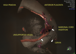
Vasa praevia is the presence of unprotected fetal blood vessels within the amniotic membranes over or near the cervical os. Rupture of the membranes before or during labour may result in fetal death from rapid blood loss if an unprotected fetal blood vessel near the cervix is damaged.
Three types are described:
Type 1 – characterised by a velamentous insertion of the umbilical cord onto the membranes distant to the placenta, with the vessels traversing the membranes over the cervix before reaching the placenta.
Type 2 – characterised by a succenturiate, multilobed or bilobed placenta with fetal vessels connecting the lobes overlaying the cervix.
Type 3 – characterised by abnormal fetal blood vessels branching off the placenta and lying unprotected near the cervical os, arising after resolution of placenta praevia or low-lying placenta.
A systematic review of 24 observational studies reported 1 case of vasa praevia for every 1,271 pregnancies (95% CI 990 to 1,692) (Zhang et al., 2023). Australian data suggest a lower rate as cited in Sullivan et al. (2017) with 63 cases from approximately 294,045 pregnancies, or 1 in 4,673. The lower Australian incidence may be explained in part by low multiple pregnancy rates arising from single embryo transfer policies (RANZCOG, 2025).
Why diagnosis matters
Screening programs for vasa praevia have been shown to be highly sensitive. When vasa praevia is not diagnosed before labour, the estimated perinatal mortality is ==44% to 70% (RANZCOG, 2025)==, and that mortality persists even with an urgent caesarean once a vessel has ruptured. The classic intrapartum presentation is painless dark-red vaginal bleeding after rupture of membranes, accompanied by acute fetal compromise. By the time this happens, the window to act is narrow.
When the diagnosis is made antenatally and a planned caesarean is performed before labour, perinatal survival is 98.6% (95% CI 96.7 to 99.7%), compared with 72.1% (95% CI 50.6 to 89.4%) where no antenatal diagnosis is made (Zhang et al., 2021). Intact perinatal survival, meaning survival without severe impairment, shows the same pattern: 96.7% with an antenatal diagnosis against 28.1% without (Zhang et al., 2021).
In short, the condition is almost entirely survivable if found in time, and frequently fatal if not. That dichotomy is what justifies screening and vigilance.
Who is at risk?
Approximately 83% of women with vasa praevia have at least one identifiable risk factor (RANZCOG, 2025). The strongest ultrasound predictors are a low-lying placenta and a velamentous cord insertion.
Pooled prevalence of each risk factor among vasa praevia cases (Pavalagantharajah et al., 2020):
Risk factor
Prevalence in vasa praevia cases (95% CI)
Low-lying placenta or resolved placenta praevia
61.5% (53.0–70.0%)
Velamentous cord insertion
52.2% (39.6–64.7%)
Bilobed or succenturiate-lobed placenta
33.3% (20.9–45.7%)
Pregnancy through assisted reproductive technology
28.2% (20.1–36.3%)
Multiple gestation
8.9% (5.3–12.5%)
Risk factors are common, and most women with them will not have vasa praevia. Velamentous cord insertion occurs in roughly 2% of pregnancies, while vasa praevia affects approximately 0.08%. Risk factors raise the index of suspicion and determine who needs a follow-up scan but they are not the diagnosis itself.
It is also worth noting that findings identified in the second trimester frequently resolve. Vasa praevia resolves in 14% to 39% of cases, with resolution far more likely when the finding is made before 24 to 26 weeks (RANZCOG, 2025). This is precisely why the diagnosis is confirmed later in pregnancy rather than applied at the morphology scan.
How is vasa praevia diagnosed?
Diagnosis is made with ultrasound, and RANZCOG describes a two-stage pathway.
Screening — 20 to 22 week morphology scan. RANZCOG suggests all women be offered screening for vasa praevia at the time of the second trimester anatomy scan (Evidence-based recommendation 1). A good quality report will comment on the location of the placental cord insertion, the morphology of the placenta, and clear documentation of a colour Doppler sweep of the cervix to assess for low-lying fetal vessels. At this stage, no distinction is drawn between vasa praevia and low-lying fetal vessels: any fetal vessels within 50 mm of the internal cervical os are characterised as low-lying fetal vessels.
Diagnosis — transvaginal ultrasound at 28 to 32 weeks. Where any of the following are present at the morphology scan, or where a woman with risk factors has not had a previous scan, the diagnosis should be confirmed by transvaginal ultrasound at 28 to 32 weeks, or earlier if clinically indicated (Evidence-based recommendation 2):
low-lying fetal vessels (within 50 mm of the cervical os)
velamentous cord insertion
low-lying placenta
bilobed or multilobed placenta
At that third trimester scan, fetal vessels within 20 mm of the cervical os identified by transvaginal ultrasound are diagnostic of vasa praevia (Evidence-based recommendation 3). Vessels between 20 mm and 50 mm are defined as low-lying fetal vessels, and vessels beyond 50 mm require no further investigation for vasa praevia.
Transvaginal ultrasound with Doppler is the preferred approach for evaluating the cervical os and lower uterine segment, providing high resolution of small vessels and a more accurate assessment of the vessel-to-os distance. Where vessels remain within 50 mm at the early third trimester scan, a repeat scan at or after 32 weeks should be considered to confirm their final location.
3D Rendered Volume with Colour Doppler
There is no international consensus on the distance that defines vasa praevia. In a 2024 survey of 57 experts, 34% used a 20 mm threshold, 32% used 50 mm, and 21% did not rely on distance at all (RANZCOG, 2025). The RANZCOG thresholds represent a pragmatic position rather than a settled one.
When to refer to a tertiary unit
For GPs and clinicians in shared-care or community settings, the referral triggers are straightforward:
A suspected or confirmed finding on ultrasound should prompt referral to specialist obstetric care for a confirmatory transvaginal scan at 28 to 32 weeks, and ongoing management planning.
Suspected vasa praevia, together with clinical features of preterm labour or antepartum haemorrhage occurring before the third-trimester confirmatory scan, warrants consultation with a Maternal Fetal Medicine (MFM) specialist for management of the pregnancy.
At the extremes of prematurity, or where the diagnosis is uncertain, MFM consultation is again advisable to balance the competing risks.
An equivocal or incomplete view of the cord insertion or the vessels near the os is reasonable grounds to refer for a closer look. The aim is simply to ensure that, where the risk exists, the vessels have been specifically assessed. When vasa praevia is excluded, that is genuine reassurance for the patient and for you. When it is identified, a clear plan can be implemented.
Delivery and management
Once vasa praevia is confirmed, patient management centres on planning a caesarean birth before the onset of labour, while weighing the risks of prematurity against the risk of vessel rupture. Care should be individualised and discussed in the context of patient preferences and individual circumstances.
For asymptomatic women with confirmed vasa praevia, inpatient versus outpatient management from 32+0 weeks should be discussed rather than mandated. Outpatient management is reasonable for asymptomatic women, particularly those without additional risk factors and with good access to emergency services. When weighing this up, consider additional risk factors for preterm labour multiple gestation, a short cervix, or a history of preterm birth. Women with low-lying fetal vessels (20 to 50 mm) can generally be managed as outpatients with ongoing antenatal care and birth planning.
Current guidance on timing of birth, balancing prematurity against the risk of vessel rupture in labour (RANZCOG, 2025):
Asymptomatic, no preterm-birth risk factors: consider caesarean from 36+0 weeks. Evidence suggests 36+0 to 36+6 weeks may best balance the risks of prematurity against those of vasa praevia complications.
Risk factors for preterm birth present: consider caesarean from 34+0 weeks.
Symptomatic (bleeding, preterm labour): consider urgent caesarean, balancing the risks of prematurity against the likelihood of fetal compromise.
Birth planning should be guided by clinical features, patient preferences, and access to specialist obstetric and neonatal services, with the aim of ensuring birth before the onset of labour or complications.
Refer your patient to QUFW
At QUFW, our consultants are subspecialist obstetricians and gynaecologists with extensive experience in high-risk pregnancies. Vasa praevia is uncommon, but primary care providers should offer a routine ultrasound at 20 to 22 weeks (morphology scan) to assess for this condition in all pregnancies.
If you would like to contact us to discuss a patient you would like assessed, or if your patient would like to book an appointment, click here.
References
Pavalagantharajah, S., Villani, L. A., & D’Souza, R. (2020). Vasa previa and associated risk factors: A systematic review and meta-analysis. American Journal of Obstetrics & Gynecology MFM, 2(3), 100117.
Sullivan, E. A., Javid, N., Duncombe, G., Li, Z., Safi, N., Cincotta, R., Homer, C. S. E., Halliday, L., & Oyelese, Y. (2017). Vasa previa diagnosis, clinical practice, and outcomes in Australia. Obstetrics & Gynecology, 130(3), 591–598.
Zhang, W., Geris, S., Al-Emara, N., Ramadan, G., Sotiriadis, A., & Akolekar, R. (2021). Perinatal outcome of pregnancies with prenatal diagnosis of vasa previa: Systematic review and meta-analysis. Ultrasound in Obstetrics & Gynecology, 57(5), 710–719.
Zhang, W., Giacchino, T., Chanyarungrojn, P. A., Ionescu, O., & Akolekar, R. (2023). Incidence of vasa praevia: A systematic review and meta-analysis. BMJ Open, 13(9), e075245.
Content Disclaimer
The information provided on this website is for educational and informational purposes only. It is not intended as a substitute for professional medical advice, diagnosis, or treatment. Always seek the advice of your obstetrician or other qualified provider for any questions you may have regarding a medical condition or treatment, and before undertaking a new healthcare regimen.
The content on this website is not intended to be a comprehensive source of information on any particular topic and should not be relied upon as such. The authors and publishers of this website are not liable for any damages or injury resulting from the use or misuse of the information provided on this website.
If you have been told you have cervical insufficiency, it is natural to want to understand what it means for the rest of your pregnancy.
Here is some reassurance: cervical insufficiency is not something you have caused, and it is not a verdict on your pregnancy. It is a recognised and manageable condition, and your team monitors it closely so they can act early if needed. With monitoring and a clear plan in place, most women in this situation go on to have a healthy baby.
This article explains what cervical insufficiency is, how it is diagnosed, the scan used to check your cervix, and what a diagnosis means for the rest of your pregnancy.
The cervix is the lower part of the uterus, the passage that connects the uterus to the vagina. For most of your pregnancy, it is meant to stay firm and closed, holding everything safely in place until your body is ready for labour.
Cervical insufficiency describes a cervix that begins to open too early, usually during the second trimester, and often without the pain or contractions you would expect with labour. It can happen quietly and can lead to a very early birth or a mid-trimester loss.
Cervical insufficiency is more common in patients who have had previous cervical surgery or biopsy (such as a cone biopsy or a LLETZ procedure), prior early pregnancy loss or previous cervical insufficiency.
How is cervical insufficiency diagnosed?
No single test diagnoses cervical insufficiency on its own. It is largely a clinical diagnosis, meaning your obstetrician puts together several pieces of information rather than relying on a single result.
Your obstetric and gynaecological history is one of the most important clues. A previous painless loss in the second trimester, or a very early birth, is the classic situation that raises the possibility of cervical insufficiency. In a current pregnancy, it may also be suspected if an examination finds that the cervix has already begun to open, or if changes are seen when your cervix is assessed on ultrasound. Where your history or risk factors suggest an increased risk, your team will usually keep a close eye on your cervix through the pregnancy so that any change is picked up early.
When is the cervix checked?
The cervix is checked and measured routinely at all pregnancy scans. Patients will be asked to present to their pregnancy scans with a comfortably full bladder. A full bladder provides an acoustic window that allows your sonographer to adequately visualise the cervix. A cervical length measuring less than 35 mm on transabdominal ultrasound will prompt your sonographer to offer a transvaginal ultrasound for a more detailed assessment. This will only be performed with your consent. A transvaginal ultrasound is the preferred and most accurate method of measuring the cervix at the morphology scan (20-22 weeks). The examination is performed with an empty bladder and minimal pressure from the transducer. A transvaginal scan lasts no more than a minute or two. Some women feel apprehensive about having a transvaginal scan in pregnancy. This technique is safe in pregnancy, and most patients find it involves little or no discomfort. When there is a reason to monitor your cervix, these scans are usually repeated at different stages of pregnancy. This is exactly the kind of careful, specialist assessment where subspecialist experience and high-end equipment make a real difference.
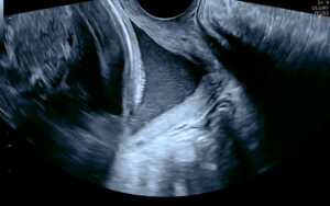
Cervical insufficiency on ultrasound
Is cervical insufficiency the same as a short cervix?
These two terms are closely related, but not the same.
It is quite normal for the cervix to shorten slightly, but remain closed, toward the end of a pregnancy. The cervix is “short” when it measures less than 25 mm on transvaginal ultrasound. The cervix can remain short and closed until the onset of labour. A short cervix is more concerning the earlier it presents in pregnancy. For most patients, it is an isolated finding that is simply monitored with ultrasound at regular intervals. Your obstetrician will interpret your situation in the context of your full history and may prescribe progesterone.
A short cervix can progress to cervical insufficiency in some cases. Cervical insufficiency is when the cervix opens prematurely, allowing the amniotic membrane to protrude into the opening. In these cases, patients are more likely to experience premature rupture of membranes (PPROM), leading to premature delivery. These patients are more at risk of infection and early pregnancy loss.
What cervical insufficiency means for the rest of your pregnancy
The good news is that cervical insufficiency is actively managed, and you will not be navigating it alone.
In practice, your care will usually include closer monitoring of your cervix during the relevant weeks of pregnancy, so that any changes are picked up early. Alongside that, there are two main treatments. One option is a cervical cerclage: a stitch placed in the cervix to give it extra support. This may be recommended in certain situations, particularly where there is a history of a previous early loss or preterm birth. Another option is vaginal progesterone, usually taken nightly. There is good evidence that supports the use of progesterone to help reduce the risk of preterm birth in people with a short cervix by supporting uterine relaxation and limiting inflammatory changes that can contribute to cervical shortening. Cervical pessaries have been used to support a short or insufficient cervix, but evidence of the benefit is inconsistent, so they are not routinely recommended for preventing preterm birth. Vaginal progesterone and, in selected cases, cervical cerclage have stronger guideline support. Your obstetrician will advise you on the most appropriate approach and intervention based on your history and your individual circumstances.
Your team may also discuss whether to adjust certain activities, and this is a conversation worth having with them directly rather than relying on general advice. The most useful thing you can do is keep the lines of communication open with your team and ask them anything that is worrying you. Closer monitoring is not a sign that something is already wrong; it is a deliberate way of staying a step ahead.
Book a cervical assessment at QUFW
At QUFW, our consultants are subspecialist obstetricians and gynaecologists with extensive experience in obstetric ultrasound, supported by highly trained sonographers. If your history means your cervix needs close monitoring, we can provide the detailed transvaginal assessment needed to evaluate it accurately and support you and your care team with a clear plan forward.
Is cervical insufficiency the same as a short cervix?
Not quite. A short cervix is a measurement found on a scan and a risk marker. Cervical insufficiency is a diagnosis describing a cervix that opens too early, usually painlessly, in the second trimester. A short cervix can point toward that risk, but the two are not the same thing.
How is cervical insufficiency diagnosed?
Mainly from your history. Particularly a previous painless second-trimester loss or very early birth. This history, together with a clinical examination and a transvaginal ultrasound of the cervix, can help your care team make a diagnosis. No single test diagnoses it on its own; your obstetrician considers the full picture.
Can you have a successful pregnancy with cervical insufficiency?
Yes. With monitoring and a clear plan. This may include a cervical stitch or progesterone, and most women go on to have a healthy baby. Cervical insufficiency increases the chance of an early birth, but it does not determine the outcome.
Does cervical insufficiency have symptoms?
Often not, which is why the cervix is checked on a scan rather than by waiting for signs. Some women may notice pelvic pressure, backache, or a change in discharge, but these are non-specific and cannot be relied on. If you have any bleeding, fluid loss, or regular tightening, contact your care team promptly.
How is cervical insufficiency treated?
The two main options are a cervical stitch (cerclage) and vaginal progesterone, chosen according to your circumstances and history, alongside closer monitoring of your cervix. Your obstetrician will recommend the approach best suited to you.
Sources: This article draws on guidance from the Royal Australian and New Zealand College of Obstetricians and Gynaecologists (RANZCOG). For more, see the RANZCOG resource hub at ranzcog.edu.au/resource-hub.
Content Disclaimer
The information provided on this website is for educational and informational purposes only. It is not intended as a substitute for professional medical advice, diagnosis, or treatment. Always seek the advice of your obstetric doctor or other qualified provider with any questions you may have about a medical condition or treatment, and before starting a new healthcare regimen.
The content on this website is not intended to be a comprehensive source of information on any particular topic and should not be relied upon as such. The authors and publishers of this website are not liable for any damages or injury resulting from the use or misuse of the information provided on this website.
For women living in remote and regional Queensland, the path to answers about pelvic pain and gynaecological issues looks very different from the one of their city counterparts. It is longer, more expensive and often means stacking a month’s worth of appointments into a single trip because coming back next week is not an option.
We recently asked one of our patients, a young woman from Western Queensland about navigating her pain and the health system while living on a farm. Her story is not unusual for women living hours from a major centre and we are grateful she was willing to share it to shine a light on the challenges facing women living in remote areas of Australia.
Lucinda at work on a cattle property in the Gulf of Carpentaria
Life on the land, and the pain that started at eleven
Home for Lucinda is Western Queensland, roughly 660 kilometres, or seven hours drive from Toowoomba. Until recently she was working as a station hand: cattle work, long days on horseback, riding motorbikes across her property.
Alongside a physically demanding job, Lucinda has lived with pain since she was eleven.
“Before my first period I remember experiencing really bad period cramps on and off and I didn’t know what it was at the time as I was 11 and hadn’t had my period before. The pain eventually got really bad, then I got my period and realised that was what the pain was.”
By thirteen or fourteen she was on the pill, prescribed to manage heavy bleeding and cramping her mother sensed was heavier than most girls her age described. She stayed on it for close to a decade because this had become the status quo for managing intense pelvic pain in women.
Lucinda at work on a cattle property
What it takes to see a specialist from 660km away
Two years ago the picture changed. She came off the pill she had been on since her early teens, and within months the pain intensified and she was back to her GP to get to the bottom of her symptoms.
What followed is the pattern many women with suspected endometriosis will recognise. Her GP raised endometriosis as a possibility, and from there the investigations added up quickly with scans, imaging, blood work, referrals to specialists and allied health practitioners across women’s health, gastroenterology and pelvic care. Each result narrowed the picture a little, but nothing definitive emerged. Along the way she was referred to QUFW for a specialised endometriosis ultrasound.
Each of those appointments posed a logistical challenge:
“I try to line up a bunch of appointments within a week or two (or four, which I had to do recently). Luckily for me I am able to stay with family in Toowoomba, because I wouldn’t be able to afford accommodation for all that time.”
Telehealth, she says, has its place, particularly for follow-ups where there is nothing physical to examine. But scans, examinations, internal assessments, and pelvic floor work cannot happen through a video link. And even for the appointments telehealth can cover, she has found that describing pelvic pain to a practitioner over a screen is harder for both sides versus being in the same room.
The real cost of travelling for care
The financial reality is difficult to overstate. Individual appointments regularly cost more than $100, some more than $200, and specialised scans can run to $500 or more. Over the past two years she has lost track of the total. Last year she reached the Medicare Safety Net threshold, a service she is grateful for, but one that only exists because the underlying spend is so high.
Beyond direct costs, the career impact has been substantial. Station work is physically demanding, and with pelvic and back pain proving difficult to treat, she had to pause this type of work.
“I have not been working for the last year or so as the work I was doing was very physically demanding and my body wasn’t keeping up. Luckily, I have been able to live with my parents, but I have had a period of 18 months where I haven’t had a steady income while trying to find a new career path that will suit my situation.”
For women without the safety nets of family, the same clinical journey would be financially unreachable.
Feeling heard, but not always understood
Lucinda said she’s “been very lucky to have a supportive medical team” and doesn’t feel like she’s been dismissed by anyone and they are mostly very quick to reinforce that her pain is real.
What she has felt is misunderstood, and deflated by the structural realities of the system.
“There are so many nuanced areas of the wider women’s health and pelvic pain field, and I have found it difficult to find practitioners that have in-depth knowledge in some of these areas. I think most of the trouble I’ve had with practitioners is not a fault of their own but is simply the fact that they are so pressured for time and where I’ve been able to get an hour-long appointment with certain practitioners, it takes almost the whole hour just to explain my pelvic pain and symptoms and the impact that its having on my life which is such a hard thing to judge when it’s about your own situation. Then you have to wait another month or two for a follow up to actually get started on an action plan. It has left me feeling pretty deflated and defeated at times.”
The scan that shaped what comes next
Recently Lucinda had a specialised endometriosis ultrasound at QUFW. Aside from her pain, nothing conclusive was found. This result is worth explaining, because it is one of the more misunderstood outcomes in this field.
An endometriosis-informed ultrasound is designed to look for deep infiltrating endometriosis and its structural signs: nodules, adhesions, endometriomas, sliding sign abnormalities, involvement of the bowel, bladder, uterosacral ligaments and pouch of Douglas. It is a much more detailed assessment than a standard pelvic ultrasound. When it does not identify these findings, it does not mean pain is not real, or that endometriosis is not present. Superficial disease in particular can sit below the resolution of any imaging modality. A thorough scan can sometimes be the cause for the next step and further investigation.
For Lucinda, the next step is a laparoscopy which is currently the only way to definitively diagnose or exclude endometriosis, and for many women this is the point where years of investigation finally gets some answers.
To other women in remote and regional Queensland
There are many women who are facing similar challenges to Lucinda, due to the complex nature of particular gynaecological issues compounded by vast geographical distances from medical care.
Keep going, find the practitioners who take the time and use resources that make sense to you.
At QUFW, we see women who have travelled across Queensland for a scan they cannot get closer to home. We know what that trip costs and it shapes how we approach every appointment.
Book an endometriosis-informed ultrasound at QUFW
If you are experiencing pelvic pain, or your treating team has raised endometriosis as a possibility, a specialised endometriosis ultrasound at QUFW is a considered next step. Our sonographers and consultants are experienced in deep infiltrating endometriosis assessment, and we work closely with your referring GP or gynaecologist to make sure the findings translate into a clear plan.
For women travelling from regional and remote Queensland, we can coordinate appointments to make the most of your time with us and you will be able to access consultations with our team of Doctors via Zoom.
The information provided on this website is for educational and informational purposes only. It is not intended as a substitute for professional medical advice, diagnosis, or treatment. Always seek the advice of your obstetric doctor or other qualified provider with any questions you may have regarding a medical condition or treatment and before undertaking a new healthcare regimen.
The content on this website is not intended to be a comprehensive source of information on any particular topic and should not be relied upon as such. The authors and publishers of this website are not liable for any damages or injury resulting from the use or misuse of the information provided on this website.
This story is shared with the patient’s permission. Some responses have been lightly edited for length and clarity.
Having preeclampsia in a previous pregnancy changes how you approach the next one. It is natural to feel a mix of hope and apprehension when you see a positive test, or even when you are still at the planning stage. What happened before stays with you, and it is completely reasonable to want answers before you find yourself back in unfamiliar territory.
The good news is that a previous diagnosis is not a verdict. It is information, and in pregnancy care, information is one of the most useful things you can have.
This fact sheet is for women who have been through preeclampsia before and explains what a subsequent pregnancy might look like. It covers the conversations to have before you conceive, through to the monitoring and management involved.
What is my actual risk of pre-eclampsia happening again?
Recurrence is possible, but it is not inevitable. Research consistently shows that the majority of women who experienced preeclampsia in a previous pregnancy do not develop it again. That said, a prior history remains one of the strongest individual risk factors, and your risk is meaningfully higher than that of someone with no history of the condition.
How high that risk is depends on several factors, including how severe your previous preeclampsia was, how early in the pregnancy it developed, whether it was early-onset (before 34 weeks) or late-onset, and whether any underlying health conditions contributed. Women who had severe or early-onset preeclampsia generally carry a higher recurrence risk than those who developed a milder form later in pregnancy.
This variation is exactly why individual risk assessment matters more than population averages. Your situation is specific to you, and understanding your personal risk profile is the foundation of good planning.
Before you conceive: conversations worth having
One of the most valuable things you can do is speak with your GP or obstetrician before you start trying to conceive. This is not always top of mind, but it can make a real difference to how your next pregnancy is managed.
The Royal Australian and New Zealand College of Obstetricians and Gynaecologists (RANZCOG) recommends that women with a history of preeclampsia be reviewed prior to a subsequent pregnancy to identify and address modifiable risk factors. This pre-conception conversation is a chance to revisit what happened in your previous pregnancy, identify any underlying conditions that may have contributed (such as hypertension, kidney disease, diabetes, or autoimmune conditions), and explore whether anything can be optimised before you fall pregnant again.
For some women, this means getting a chronic condition better controlled. For others, it may simply mean having an early referral pathway in place so that care begins promptly from the moment a pregnancy is confirmed. Either way, starting that conversation early puts you in a stronger position.
How does risk screening differ in my next pregnancy?
The first trimester combined screening for preeclampsia, typically performed at your 13 to 14 week scan, still applies in a subsequent pregnancy and remains a valuable tool. What changes is the weight your history carries within that screening.
A prior history of preeclampsia is factored into the risk calculation alongside your blood pressure readings, blood test markers (including PAPP-A and PlGF), and uterine artery Doppler measurements. Because your previous experience is already a significant risk factor, you may be more likely to receive a high-risk result than someone without that history. This is not the screening working against you. It is the screening working as intended, by accounting for your full picture rather than treating you as a first-time case.
Ultrasound of the blood flow through a uterine artery (Transabdominal assessment of the uterine artery Doppler waveform in the first trimester)
It is also worth understanding that a high-risk result is not a diagnosis. Many women who screen as high-risk go on to have healthy pregnancies without developing preeclampsia. The purpose of early screening is to personalise your care and enable timely intervention where it is needed.
For more detail on what the screening involves and what a high-risk result means, see our article.
Is Aspirin suitable for preeclampsia?
If you have a prior history of preeclampsia, low-dose aspirin will very likely be recommended as a preventative measure in your next pregnancy. For women who have been through peeclampsia before, the reasoning behind this recommendation often lands differently. You already know what it is trying to prevent.
RANZCOG and the Society of Obstetric Medicine of Australia and New Zealand (SOMANZ) both support the use of low-dose aspirin for women at increased risk of preeclampsia. The recommended dose is 150mg taken at night, commenced between 11–14 weeks of pregnancy and continued until 34 to 36 weeks gestation.
Evidence from the ASPRE trial shows that starting aspirin at this dose and timing can reduce the risk of preterm preeclampsia (before 37 weeks) by approximately 62%, and early preeclampsia (before 34 weeks) by approximately 82%. It will not eliminate risk entirely, but it is one of the most effective tools currently available.
Aspirin is considered safe in pregnancy for most women, but always confirm with your treating doctor before commencing, particularly if your circumstances have changed since your last pregnancy, or if you have any known allergies or medical contraindications.
What does a preeclampsia monitoring plan might look like?
Women with a prior history of preeclampsia are typically managed as higher-risk from the outset of a new pregnancy. In practical terms, this means your care will likely include more touchpoints than a standard low-risk pregnancy.
You can expect some or all of the following, depending on your individual circumstances and your treating team’s recommendations: earlier referral to an obstetrician or maternal-fetal medicine specialist, regular blood pressure monitoring throughout the pregnancy, urine testing for protein at antenatal appointments, blood tests to monitor kidney and liver function, additional growth scans in the second and third trimester to assess your baby’s development, and placental function assessment, including Doppler studies, particularly if growth concerns arise.
This level of monitoring can feel intensive, and it is worth acknowledging that more appointments and more testing can themselves become a source of stress. It helps to reframe closer monitoring not as a sign that something is already wrong, but as a deliberate strategy to catch any changes early, when options are broader and outcomes are generally better.
What happens if I get preeclampsia again?
For some women, preeclampsia will develop again despite best efforts at prevention and monitoring. If that happens, earlier detection usually means more options and a more managed response.
Having been through it before, you are also better equipped than most. You are more likely to recognise early symptoms, more likely to know when to escalate a concern, and more likely to have a care team who is already prepared and monitoring closely. None of that eliminates the difficulty, but it does change the experience in meaningful ways.
SOMANZ guidance emphasises that women with recurrent preeclampsia benefit from specialist involvement early in pregnancy, and that a clearly documented management plan, established before symptoms develop, leads to better outcomes for both mother and baby.
Book your 13-week scan at QUFW
At QUFW, preeclampsia risk screening is included as part of our routine 13 to 14 week scan. If you have a history of preeclampsia, this appointment is an important early step in understanding your risk in this pregnancy.
Our team will take into account your full history as part of the screening process. We screen all women at this stage, and we are experienced in supporting those who have been through preeclampsia before.
The information provided on this website is for educational and informational purposes only. It is not intended as a substitute for professional medical advice, diagnosis, or treatment. Always seek the advice of your obstetric doctor or other qualified provider with any questions you may have regarding a medical condition or treatment and before undertaking a new healthcare regimen.
The content on this website is not intended to be a comprehensive source of information on any particular topic and should not be relied upon as such. The authors and publishers of this website are not liable for any damages or injury resulting from the use or misuse of the information provided on this website.
Fertility investigations can feel overwhelming, particularly when a test is new or unfamiliar. HyFoSy is a straightforward ultrasound procedure, and it’s common for patients to have questions about comfort, what will happen on the day, and how the results may guide next steps in care.
This fact sheet explains what a HyFoSy examination is, why it is performed, and what you can expect before, during, and after your appointment. In this article, Jacqui, Lead Gynaecological Sonographer at QUFW, talks you through the HyFoSy procedure and how it fits into a fertility assessment.
Medical disclaimer: The information provided here is general in nature and intended for educational purposes only. It does not replace medical advice, diagnosis, or treatment. Please speak with your GP or specialist for personalised guidance.
HyFoSy (Hysterosalpingo-Foam Sonography) is a specialised gynaecology ultrasound used as part of a fertility assessment. It evaluates the internal cavity of the uterus and the openness of the fallopian tubes.
“HyFoSy is a specialised gynaecology ultrasound procedure used as part of a fertility assessment,” Jacqui explains in the above video.
The procedure uses a contrast agent called ExEm foam, which is made from a gel and purified water. When mixed, it forms a foam that can be seen clearly on ultrasound.
“It’s used to detect any blockages in the fallopian tubes.”
Unlike X-ray–based tests, HyFoSy is performed entirely under ultrasound guidance and does not involve radiation.
Why does fallopian tube patency matter?
The fallopian tubes play a central role in natural conception.
“The fallopian tubes are an essential part of a woman’s reproductive system, connecting the ovaries to the uterus,” says Jacqui.
For pregnancy to occur:
The ovary must release an egg
The egg must travel through an open fallopian tube
Sperm must meet the egg within the tube
“If both tubes are blocked, the sperm and egg cannot meet, making conception impossible.”
Because fallopian tubes cannot be visualised on a standard pelvic ultrasound, a HyFoSy is used to assess whether they are open (patent).
When is a HyFoSy performed?
Timing is important for safety and accuracy.
“The procedure should be performed between four to ten days after the first day of your last menstrual period,” Jacqui explains.
This timing:
Reduces the chance of early pregnancy
Provides optimal visualisation
Our doctors perform the procedure after a thorough transvaginal assessment of the pelvic organs has been performed by our sonographers.
Patients are advised to contact QUFW on day one of their period to arrange an appointment. If cycles are irregular or infrequent, individual advice is provided.
How do I prepare for a HyFoSy?
Preparation is straightforward and designed to support comfort.
“You do not need a full bladder for the examination,” Jacqui notes.
You may be advised to:
Avoid sexual intercourse before the procedure
Have a pregnancy blood test if required
Take your usual pain relief one hour before the appointment
You are welcome to bring a support person if that feels helpful.
What happens during a HyFoSy?
On arrival, you will complete a urine pregnancy test to confirm you are not pregnant.
A transvaginal ultrasound is performed first to assess the uterus and ovaries. Once this is complete, the doctor will:
Insert a speculum
Place a thin, flexible catheter through the cervix into the uterus
“As the catheter passes through, you may feel some discomfort or cramping,” Jacqui explains.
A small balloon at the end of the catheter is inflated to keep it in place, which may cause brief pressure.
The speculum is then removed, and the transvaginal probe is reinserted. Saline is passed through the catheter to assess the uterine cavity for scarring, fibroids, or polyps.
Then, the ExEm foam is introduced.
“The goal is to see the contrast flow through open tubes and spill into the area surrounding the ovaries.”
Once complete, the catheter and probe are removed.
What will I feel during a HyFoSy?
Experiences vary between patients.
“Some patients may feel light-headed, hot, or dizzy during the procedure,” says Jacqui.
Common sensations include:
Mild to moderate cramping
Period-like discomfort
Temporary pressure
“These symptoms usually settle quickly,” Jacqui reassures.
Patients are encouraged to communicate during the procedure so comfort can be supported.
How long does a HyFoSy appointment take?
“We allow 45 to 60 minutes for the appointment,” Jacqui explains.
The actual HyFoSy procedure typically takes around 10 minutes, with the remainder of the time used for preparation, explanation, and ultrasound assessment.
When and how do I receive HyFoSy results?
As the procedure is performed, findings are usually discussed in real time.
“The doctor will explain the findings and whether the tubes appear open.”
Occasionally, results may be inconclusive due to temporary tubal spasm rather than true blockage.
Your results are:
Sent to your referring doctor
Shared with you via a secure Tricefy link on your mobile
Report timing depends on examination complexity and additional findings.
Is HyFoSy safe and accurate?
HyFoSy is considered a safe and well-tolerated procedure. It does not involve ionising radiation or general anaesthetic.
Studies have shown HyFoSy to have high diagnostic accuracy for tubal patency, comparable to traditional hysterosalpingography (HSG), with better patient comfort and fewer risks.
After the procedure: recovery and care
After a HyFoSy, it is normal to experience:
Watery discharge
Light bleeding or spotting
Mild cramping or bloating
“This is normal and due to the saline and contrast used,” Jacqui explains.
A sanitary pad is provided, and symptoms usually resolve within a day or two. Normal activities, including intercourse, can be resumed.
If you experience:
Severe pelvic pain
Fever
Green or yellow vaginal discharge
you should contact your GP or referring doctor, as these may indicate infection, which is uncommon.
Book your gynaecology scan at QUFW
Fertility investigations can bring up many emotions, including hope, worry, and uncertainty. Having clear information can make the process feel more manageable.
At QUFW, gynaecology scans are performed in a supportive environment using evidence-based techniques and clear communication. Your HyFoSy examination is designed to provide meaningful information to guide your fertility care, while prioritising safety and comfort.
You are supported every step of the way.
References
Exacoustos, C., Di Giovanni, A., Szabolcs, B. et al. (2015). Automated sonographic tubal patency evaluation using contrast agent (HyFoSy). Ultrasound in Obstetrics & Gynaecology, 46(5), 620–627. https://pubmed.ncbi.nlm.nih.gov/19852043/
Dreyer, K., Out, R., Hompes, P. G. et al. (2017). Oil-based or water-based contrast for hysterosalpingography in infertile women. New England Journal of Medicine, 376, 2043–2052. https://www.nejm.org/doi/full/10.1056/NEJMoa1612337
Video transcript
Hi, my name’s Jacqui. I’m the lead gynaecological sonographer for QUFW. I’m here to talk you through your HyFoSy examination at QUFW.
HyFoSy is a specialised gynaecology ultrasound procedure used as part of a fertility assessment. It provides information about the internal cavity of the uterus and the fallopian tubes. A contrast agent, ExEm foam, is used to visualise the tubes. It is made from a gel and purified water which, when mixed, create a foam used to detect any blockages in the fallopian tubes.
Why are patent fallopian tubes important?
The fallopian tubes are an essential part of a woman’s reproductive system, connecting the ovaries to the uterus. Open (or patent) fallopian tubes are important for conception. If both tubes are blocked, the sperm and egg cannot meet, making conception impossible. The fallopian tubes cannot be visualised on a standard ultrasound, which is why the HyFoSy procedure is used.
When the procedure is performed
The procedure should be performed between four to ten days after the first day of your last menstrual period. We recommend calling our office on day one of your period to make a booking. If you have irregular or infrequent cycles, please call for individual advice.
Some patients may be asked to avoid sexual intercourse before the procedure and/or have a pregnancy blood test. You do not need a full bladder for the examination. It’s recommended that you take your usual pain relief about one hour before your appointment. You may bring a support person if you wish, although this is not required.
What happens on the day
On arrival, you’ll be asked to perform a urine pregnancy test to confirm you are not pregnant. A transvaginal ultrasound is done first. Once this is complete, the doctor will insert a speculum and place a thin, flexible catheter through your cervix into the uterus. As the catheter passes through, you may feel some discomfort or cramping. Once it’s in position, a small balloon at the end of the catheter is inflated, which may cause mild pressure that should ease quickly.
The speculum is then removed and the transvaginal probe reinserted so we can visualise the procedure. Saline (a sterile saltwater solution) is passed through the catheter to check the uterine cavity for scarring, polyps, or fibroids that may not be visible on regular ultrasound. Then, the contrast agent (ExEm foam) is passed through the catheter. The doctor observes the contrast as it moves through the uterine cavity and fallopian tubes. The goal is to see the contrast flow through open tubes and spill into the area surrounding the ovaries.
When the procedure is finished, the catheter and probe are removed.
What to expect during a HyFoSy
Some patients may feel light-headed, hot, or dizzy during the procedure. Mild cramping, similar to period pain, is also common. These symptoms usually settle quickly, and pain relief can help if needed. Please tell your doctor or sonographer if you feel unwell, and they’ll help you feel more comfortable.
How long it takes
We allow 45 to 60 minutes for the appointment, including preparation and scanning time. The actual procedure usually takes about 10 minutes.
When you’ll receive your results
As the procedure is performed, the doctor will explain the findings and whether the tubes appear open. Occasionally, results may be inconclusive if it’s difficult to distinguish between tubal spasm and blockage.
Your results will be sent directly to your referring doctor, and you’ll also receive a copy via a secure link through Tricefy on your mobile. The time taken for your doctor to receive the written report will depend on the complexity of the examination and whether additional findings were noted.
After the procedure
You may notice some watery or light bloody discharge afterward. This is normal and due to the saline and contrast used. A sanitary pad will be provided. Mild bloating, cramping, or lower abdominal discomfort can occur and is usually relieved with standard pain medication. Light bleeding or spotting may continue for a day or two.
If you experience severe pelvic pain, fever, or an unusual vaginal discharge that is green or yellow, contact your GP or referring doctor promptly, as these may be signs of infection (which is uncommon).
It is safe to resume normal activities, including intercourse, after the procedure. The HyFoSy is a very safe procedure that does not involve ionising radiation or general anaesthetic.
The information provided on this website is for educational and informational purposes only. It is not intended as a substitute for professional medical advice, diagnosis, or treatment. Always seek the advice of your obstetric doctor or other qualified provider with any questions you may have regarding a medical condition or treatment and before undertaking a new healthcare regimen.
The content on this website is not intended to be a comprehensive source of information on any particular topic and should not be relied upon as such. The authors and publishers of this website are not liable for any damages or injury resulting from the use or misuse of the information provided on this website.