by Ben Sweetlove | Sep 15, 2026 | Pregnancy, QUFW News
Vasa praevia is a near-perfect example of a condition where ultrasound can genuinely change a pregnancy’s outcome. Vasa praevia can be silent; the baby gives no warning, and the only thing standing between a safe, planned birth and a preventable tragedy is early diagnosis in experienced hands.
Patients diagnosed with vasa praevia have emphasised the importance of receiving clear, evidence-based information about the risks and benefits of different care pathways. The primary benefit of screening for vasa praevia is that antenatal diagnosis significantly increases the likelihood of perinatal survival.
This post summarises current best practice, drawing on the RANZCOG guideline Vasa Praevia (C-Obs 47), Version 3 (July 2025), the most relevant evidence-based resource for practice in Australia and Aotearoa New Zealand. Written for referring GPs and clinicians, it covers what vasa praevia is, why it matters so much at delivery, the histories and findings that should raise your index of suspicion, and when targeted specialist assessment is worth arranging. (You may also see it written as vasa previa, the spelling used in much of the international literature; the two terms describe the same condition.)
Table of contents
- What is vasa praevia?
- Why diagnosis matters
- Who is at risk?
- How is vasa praevia diagnosed?
- When to refer to a tertiary unit
- Delivery and management
- Refer your patient to QUFW
What is vasa praevia?
Vasa praevia is the presence of unprotected fetal blood vessels within the amniotic membranes over or near the cervical os. Rupture of the membranes before or during labour may result in fetal death from rapid blood loss if an unprotected fetal blood vessel near the cervix is damaged.
Three types are described:
- Type 1 – characterised by a velamentous insertion of the umbilical cord onto the membranes distant to the placenta, with the vessels traversing the membranes over the cervix before reaching the placenta.
- Type 2 – characterised by a succenturiate, multilobed or bilobed placenta with fetal vessels connecting the lobes overlaying the cervix.
- Type 3 – characterised by abnormal fetal blood vessels branching off the placenta and lying unprotected near the cervical os, arising after resolution of placenta praevia or low-lying placenta.
A systematic review of 24 observational studies reported 1 case of vasa praevia for every 1,271 pregnancies (95% CI 990 to 1,692) (Zhang et al., 2023). Australian data suggest a lower rate as cited in Sullivan et al. (2017) with 63 cases from approximately 294,045 pregnancies, or 1 in 4,673. The lower Australian incidence may be explained in part by low multiple pregnancy rates arising from single embryo transfer policies (RANZCOG, 2025).
Why diagnosis matters
Screening programs for vasa praevia have been shown to be highly sensitive. When vasa praevia is not diagnosed before labour, the estimated perinatal mortality is ==44% to 70% (RANZCOG, 2025)==, and that mortality persists even with an urgent caesarean once a vessel has ruptured. The classic intrapartum presentation is painless dark-red vaginal bleeding after rupture of membranes, accompanied by acute fetal compromise. By the time this happens, the window to act is narrow.
When the diagnosis is made antenatally and a planned caesarean is performed before labour, perinatal survival is 98.6% (95% CI 96.7 to 99.7%), compared with 72.1% (95% CI 50.6 to 89.4%) where no antenatal diagnosis is made (Zhang et al., 2021). Intact perinatal survival, meaning survival without severe impairment, shows the same pattern: 96.7% with an antenatal diagnosis against 28.1% without (Zhang et al., 2021).
In short, the condition is almost entirely survivable if found in time, and frequently fatal if not. That dichotomy is what justifies screening and vigilance.
Who is at risk?
Approximately 83% of women with vasa praevia have at least one identifiable risk factor (RANZCOG, 2025). The strongest ultrasound predictors are a low-lying placenta and a velamentous cord insertion.
Pooled prevalence of each risk factor among vasa praevia cases (Pavalagantharajah et al., 2020):
| Risk factor |
Prevalence in vasa praevia cases (95% CI) |
| Low-lying placenta or resolved placenta praevia |
61.5% (53.0–70.0%) |
| Velamentous cord insertion |
52.2% (39.6–64.7%) |
| Bilobed or succenturiate-lobed placenta |
33.3% (20.9–45.7%) |
| Pregnancy through assisted reproductive technology |
28.2% (20.1–36.3%) |
| Multiple gestation |
8.9% (5.3–12.5%) |
Risk factors are common, and most women with them will not have vasa praevia. Velamentous cord insertion occurs in roughly 2% of pregnancies, while vasa praevia affects approximately 0.08%. Risk factors raise the index of suspicion and determine who needs a follow-up scan but they are not the diagnosis itself.
It is also worth noting that findings identified in the second trimester frequently resolve. Vasa praevia resolves in 14% to 39% of cases, with resolution far more likely when the finding is made before 24 to 26 weeks (RANZCOG, 2025). This is precisely why the diagnosis is confirmed later in pregnancy rather than applied at the morphology scan.
How is vasa praevia diagnosed?
Diagnosis is made with ultrasound, and RANZCOG describes a two-stage pathway.
Screening — 20 to 22 week morphology scan. RANZCOG suggests all women be offered screening for vasa praevia at the time of the second trimester anatomy scan (Evidence-based recommendation 1). A good quality report will comment on the location of the placental cord insertion, the morphology of the placenta, and clear documentation of a colour Doppler sweep of the cervix to assess for low-lying fetal vessels. At this stage, no distinction is drawn between vasa praevia and low-lying fetal vessels: any fetal vessels within 50 mm of the internal cervical os are characterised as low-lying fetal vessels.
Diagnosis — transvaginal ultrasound at 28 to 32 weeks. Where any of the following are present at the morphology scan, or where a woman with risk factors has not had a previous scan, the diagnosis should be confirmed by transvaginal ultrasound at 28 to 32 weeks, or earlier if clinically indicated (Evidence-based recommendation 2):
- low-lying fetal vessels (within 50 mm of the cervical os)
- velamentous cord insertion
- low-lying placenta
- bilobed or multilobed placenta
At that third trimester scan, fetal vessels within 20 mm of the cervical os identified by transvaginal ultrasound are diagnostic of vasa praevia (Evidence-based recommendation 3). Vessels between 20 mm and 50 mm are defined as low-lying fetal vessels, and vessels beyond 50 mm require no further investigation for vasa praevia.
Transvaginal ultrasound with Doppler is the preferred approach for evaluating the cervical os and lower uterine segment, providing high resolution of small vessels and a more accurate assessment of the vessel-to-os distance. Where vessels remain within 50 mm at the early third trimester scan, a repeat scan at or after 32 weeks should be considered to confirm their final location.
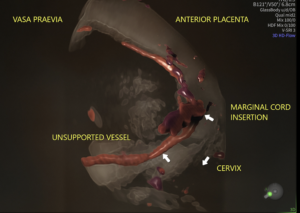
3D Rendered Volume with Colour Doppler
There is no international consensus on the distance that defines vasa praevia. In a 2024 survey of 57 experts, 34% used a 20 mm threshold, 32% used 50 mm, and 21% did not rely on distance at all (RANZCOG, 2025). The RANZCOG thresholds represent a pragmatic position rather than a settled one.
When to refer to a tertiary unit
For GPs and clinicians in shared-care or community settings, the referral triggers are straightforward:
- A suspected or confirmed finding on ultrasound should prompt referral to specialist obstetric care for a confirmatory transvaginal scan at 28 to 32 weeks, and ongoing management planning.
- Suspected vasa praevia, together with clinical features of preterm labour or antepartum haemorrhage occurring before the third-trimester confirmatory scan, warrants consultation with a Maternal Fetal Medicine (MFM) specialist for management of the pregnancy.
- At the extremes of prematurity, or where the diagnosis is uncertain, MFM consultation is again advisable to balance the competing risks.
An equivocal or incomplete view of the cord insertion or the vessels near the os is reasonable grounds to refer for a closer look. The aim is simply to ensure that, where the risk exists, the vessels have been specifically assessed. When vasa praevia is excluded, that is genuine reassurance for the patient and for you. When it is identified, a clear plan can be implemented.
Delivery and management
Once vasa praevia is confirmed, patient management centres on planning a caesarean birth before the onset of labour, while weighing the risks of prematurity against the risk of vessel rupture. Care should be individualised and discussed in the context of patient preferences and individual circumstances.
For asymptomatic women with confirmed vasa praevia, inpatient versus outpatient management from 32+0 weeks should be discussed rather than mandated. Outpatient management is reasonable for asymptomatic women, particularly those without additional risk factors and with good access to emergency services. When weighing this up, consider additional risk factors for preterm labour multiple gestation, a short cervix, or a history of preterm birth. Women with low-lying fetal vessels (20 to 50 mm) can generally be managed as outpatients with ongoing antenatal care and birth planning.
Current guidance on timing of birth, balancing prematurity against the risk of vessel rupture in labour (RANZCOG, 2025):
- Asymptomatic, no preterm-birth risk factors: consider caesarean from 36+0 weeks. Evidence suggests 36+0 to 36+6 weeks may best balance the risks of prematurity against those of vasa praevia complications.
- Risk factors for preterm birth present: consider caesarean from 34+0 weeks.
- Symptomatic (bleeding, preterm labour): consider urgent caesarean, balancing the risks of prematurity against the likelihood of fetal compromise.
Birth planning should be guided by clinical features, patient preferences, and access to specialist obstetric and neonatal services, with the aim of ensuring birth before the onset of labour or complications.
Refer your patient to QUFW
At QUFW, our consultants are subspecialist obstetricians and gynaecologists with extensive experience in high-risk pregnancies. Vasa praevia is uncommon, but primary care providers should offer a routine ultrasound at 20 to 22 weeks (morphology scan) to assess for this condition in all pregnancies.
If you would like to contact us to discuss a patient you would like assessed, or if your patient would like to book an appointment, click here.
References
Pavalagantharajah, S., Villani, L. A., & D’Souza, R. (2020). Vasa previa and associated risk factors: A systematic review and meta-analysis. American Journal of Obstetrics & Gynecology MFM, 2(3), 100117.
Royal Australian and New Zealand College of Obstetricians and Gynaecologists. (2025). Vasa praevia (C-Obs 47, Clinical Guideline Version 3).
Sullivan, E. A., Javid, N., Duncombe, G., Li, Z., Safi, N., Cincotta, R., Homer, C. S. E., Halliday, L., & Oyelese, Y. (2017). Vasa previa diagnosis, clinical practice, and outcomes in Australia. Obstetrics & Gynecology, 130(3), 591–598.
Zhang, W., Geris, S., Al-Emara, N., Ramadan, G., Sotiriadis, A., & Akolekar, R. (2021). Perinatal outcome of pregnancies with prenatal diagnosis of vasa previa: Systematic review and meta-analysis. Ultrasound in Obstetrics & Gynecology, 57(5), 710–719.
Zhang, W., Giacchino, T., Chanyarungrojn, P. A., Ionescu, O., & Akolekar, R. (2023). Incidence of vasa praevia: A systematic review and meta-analysis. BMJ Open, 13(9), e075245.
Content Disclaimer
The information provided on this website is for educational and informational purposes only. It is not intended as a substitute for professional medical advice, diagnosis, or treatment. Always seek the advice of your obstetrician or other qualified provider for any questions you may have regarding a medical condition or treatment, and before undertaking a new healthcare regimen.
The content on this website is not intended to be a comprehensive source of information on any particular topic and should not be relied upon as such. The authors and publishers of this website are not liable for any damages or injury resulting from the use or misuse of the information provided on this website.
by Ben Sweetlove | Sep 15, 2026 | Pregnancy, QUFW News
If you have been told you have cervical insufficiency, it is natural to want to understand what it means for the rest of your pregnancy.
Here is some reassurance: cervical insufficiency is not something you have caused, and it is not a verdict on your pregnancy. It is a recognised and manageable condition, and your team monitors it closely so they can act early if needed. With monitoring and a clear plan in place, most women in this situation go on to have a healthy baby.
This article explains what cervical insufficiency is, how it is diagnosed, the scan used to check your cervix, and what a diagnosis means for the rest of your pregnancy.
Table of contents
- What is cervical insufficiency?
- How is cervical insufficiency diagnosed?
- The scan used to check your cervix
- Is cervical insufficiency the same as a short cervix?
- What cervical insufficiency means for the rest of your pregnancy
- Book a cervical assessment at QUFW
- Frequently asked questions
What is cervical insufficiency?
The cervix is the lower part of the uterus, the passage that connects the uterus to the vagina. For most of your pregnancy, it is meant to stay firm and closed, holding everything safely in place until your body is ready for labour.
Cervical insufficiency describes a cervix that begins to open too early, usually during the second trimester, and often without the pain or contractions you would expect with labour. It can happen quietly and can lead to a very early birth or a mid-trimester loss.
Cervical insufficiency is more common in patients who have had previous cervical surgery or biopsy (such as a cone biopsy or a LLETZ procedure), prior early pregnancy loss or previous cervical insufficiency.
How is cervical insufficiency diagnosed?
No single test diagnoses cervical insufficiency on its own. It is largely a clinical diagnosis, meaning your obstetrician puts together several pieces of information rather than relying on a single result.
Your obstetric and gynaecological history is one of the most important clues. A previous painless loss in the second trimester, or a very early birth, is the classic situation that raises the possibility of cervical insufficiency. In a current pregnancy, it may also be suspected if an examination finds that the cervix has already begun to open, or if changes are seen when your cervix is assessed on ultrasound. Where your history or risk factors suggest an increased risk, your team will usually keep a close eye on your cervix through the pregnancy so that any change is picked up early.
When is the cervix checked?
The cervix is checked and measured routinely at all pregnancy scans. Patients will be asked to present to their pregnancy scans with a comfortably full bladder. A full bladder provides an acoustic window that allows your sonographer to adequately visualise the cervix. A cervical length measuring less than 35 mm on transabdominal ultrasound will prompt your sonographer to offer a transvaginal ultrasound for a more detailed assessment. This will only be performed with your consent. A transvaginal ultrasound is the preferred and most accurate method of measuring the cervix at the morphology scan (20-22 weeks). The examination is performed with an empty bladder and minimal pressure from the transducer. A transvaginal scan lasts no more than a minute or two. Some women feel apprehensive about having a transvaginal scan in pregnancy. This technique is safe in pregnancy, and most patients find it involves little or no discomfort. When there is a reason to monitor your cervix, these scans are usually repeated at different stages of pregnancy. This is exactly the kind of careful, specialist assessment where subspecialist experience and high-end equipment make a real difference.
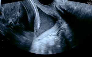
Cervical insufficiency on ultrasound
Is cervical insufficiency the same as a short cervix?
These two terms are closely related, but not the same.
It is quite normal for the cervix to shorten slightly, but remain closed, toward the end of a pregnancy. The cervix is “short” when it measures less than 25 mm on transvaginal ultrasound. The cervix can remain short and closed until the onset of labour. A short cervix is more concerning the earlier it presents in pregnancy. For most patients, it is an isolated finding that is simply monitored with ultrasound at regular intervals. Your obstetrician will interpret your situation in the context of your full history and may prescribe progesterone.
A short cervix can progress to cervical insufficiency in some cases. Cervical insufficiency is when the cervix opens prematurely, allowing the amniotic membrane to protrude into the opening. In these cases, patients are more likely to experience premature rupture of membranes (PPROM), leading to premature delivery. These patients are more at risk of infection and early pregnancy loss.
What cervical insufficiency means for the rest of your pregnancy
The good news is that cervical insufficiency is actively managed, and you will not be navigating it alone.
In practice, your care will usually include closer monitoring of your cervix during the relevant weeks of pregnancy, so that any changes are picked up early. Alongside that, there are two main treatments. One option is a cervical cerclage: a stitch placed in the cervix to give it extra support. This may be recommended in certain situations, particularly where there is a history of a previous early loss or preterm birth. Another option is vaginal progesterone, usually taken nightly. There is good evidence that supports the use of progesterone to help reduce the risk of preterm birth in people with a short cervix by supporting uterine relaxation and limiting inflammatory changes that can contribute to cervical shortening. Cervical pessaries have been used to support a short or insufficient cervix, but evidence of the benefit is inconsistent, so they are not routinely recommended for preventing preterm birth. Vaginal progesterone and, in selected cases, cervical cerclage have stronger guideline support. Your obstetrician will advise you on the most appropriate approach and intervention based on your history and your individual circumstances.
Your team may also discuss whether to adjust certain activities, and this is a conversation worth having with them directly rather than relying on general advice. The most useful thing you can do is keep the lines of communication open with your team and ask them anything that is worrying you. Closer monitoring is not a sign that something is already wrong; it is a deliberate way of staying a step ahead.
Book a cervical assessment at QUFW
At QUFW, our consultants are subspecialist obstetricians and gynaecologists with extensive experience in obstetric ultrasound, supported by highly trained sonographers. If your history means your cervix needs close monitoring, we can provide the detailed transvaginal assessment needed to evaluate it accurately and support you and your care team with a clear plan forward.
If you would like to arrange an assessment, click here to book an appointment. We are here for you.
Frequently asked questions
Is cervical insufficiency the same as a short cervix?
Not quite. A short cervix is a measurement found on a scan and a risk marker. Cervical insufficiency is a diagnosis describing a cervix that opens too early, usually painlessly, in the second trimester. A short cervix can point toward that risk, but the two are not the same thing.
How is cervical insufficiency diagnosed?
Mainly from your history. Particularly a previous painless second-trimester loss or very early birth. This history, together with a clinical examination and a transvaginal ultrasound of the cervix, can help your care team make a diagnosis. No single test diagnoses it on its own; your obstetrician considers the full picture.
Can you have a successful pregnancy with cervical insufficiency?
Yes. With monitoring and a clear plan. This may include a cervical stitch or progesterone, and most women go on to have a healthy baby. Cervical insufficiency increases the chance of an early birth, but it does not determine the outcome.
Does cervical insufficiency have symptoms?
Often not, which is why the cervix is checked on a scan rather than by waiting for signs. Some women may notice pelvic pressure, backache, or a change in discharge, but these are non-specific and cannot be relied on. If you have any bleeding, fluid loss, or regular tightening, contact your care team promptly.
How is cervical insufficiency treated?
The two main options are a cervical stitch (cerclage) and vaginal progesterone, chosen according to your circumstances and history, alongside closer monitoring of your cervix. Your obstetrician will recommend the approach best suited to you.
Sources: This article draws on guidance from the Royal Australian and New Zealand College of Obstetricians and Gynaecologists (RANZCOG). For more, see the RANZCOG resource hub at ranzcog.edu.au/resource-hub.
Content Disclaimer
The information provided on this website is for educational and informational purposes only. It is not intended as a substitute for professional medical advice, diagnosis, or treatment. Always seek the advice of your obstetric doctor or other qualified provider with any questions you may have about a medical condition or treatment, and before starting a new healthcare regimen.
The content on this website is not intended to be a comprehensive source of information on any particular topic and should not be relied upon as such. The authors and publishers of this website are not liable for any damages or injury resulting from the use or misuse of the information provided on this website.
by Ben Sweetlove | Apr 23, 2026 | Pregnancy, QUFW News
Having preeclampsia in a previous pregnancy changes how you approach the next one. It is natural to feel a mix of hope and apprehension when you see a positive test, or even when you are still at the planning stage. What happened before stays with you, and it is completely reasonable to want answers before you find yourself back in unfamiliar territory.
The good news is that a previous diagnosis is not a verdict. It is information, and in pregnancy care, information is one of the most useful things you can have.
This fact sheet is for women who have been through preeclampsia before and explains what a subsequent pregnancy might look like. It covers the conversations to have before you conceive, through to the monitoring and management involved.
Table of contents
- What is my actual risk of it happening again?
- Before you conceive: conversations worth having
- How risk screening differs in your next pregnancy
- Aspirin: earlier, and with more context this time
- What a monitoring plan might look like
- If preeclampsia does recur
- Book your 13-week scan at QUFW
What is my actual risk of pre-eclampsia happening again?
Recurrence is possible, but it is not inevitable. Research consistently shows that the majority of women who experienced preeclampsia in a previous pregnancy do not develop it again. That said, a prior history remains one of the strongest individual risk factors, and your risk is meaningfully higher than that of someone with no history of the condition.
How high that risk is depends on several factors, including how severe your previous preeclampsia was, how early in the pregnancy it developed, whether it was early-onset (before 34 weeks) or late-onset, and whether any underlying health conditions contributed. Women who had severe or early-onset preeclampsia generally carry a higher recurrence risk than those who developed a milder form later in pregnancy.
This variation is exactly why individual risk assessment matters more than population averages. Your situation is specific to you, and understanding your personal risk profile is the foundation of good planning.
Before you conceive: conversations worth having
One of the most valuable things you can do is speak with your GP or obstetrician before you start trying to conceive. This is not always top of mind, but it can make a real difference to how your next pregnancy is managed.
The Royal Australian and New Zealand College of Obstetricians and Gynaecologists (RANZCOG) recommends that women with a history of preeclampsia be reviewed prior to a subsequent pregnancy to identify and address modifiable risk factors. This pre-conception conversation is a chance to revisit what happened in your previous pregnancy, identify any underlying conditions that may have contributed (such as hypertension, kidney disease, diabetes, or autoimmune conditions), and explore whether anything can be optimised before you fall pregnant again.
For some women, this means getting a chronic condition better controlled. For others, it may simply mean having an early referral pathway in place so that care begins promptly from the moment a pregnancy is confirmed. Either way, starting that conversation early puts you in a stronger position.
How does risk screening differ in my next pregnancy?
The first trimester combined screening for preeclampsia, typically performed at your 13 to 14 week scan, still applies in a subsequent pregnancy and remains a valuable tool. What changes is the weight your history carries within that screening.
A prior history of preeclampsia is factored into the risk calculation alongside your blood pressure readings, blood test markers (including PAPP-A and PlGF), and uterine artery Doppler measurements. Because your previous experience is already a significant risk factor, you may be more likely to receive a high-risk result than someone without that history. This is not the screening working against you. It is the screening working as intended, by accounting for your full picture rather than treating you as a first-time case.
Ultrasound of the blood flow through a uterine artery (Transabdominal assessment of the uterine artery Doppler waveform in the first trimester)
It is also worth understanding that a high-risk result is not a diagnosis. Many women who screen as high-risk go on to have healthy pregnancies without developing preeclampsia. The purpose of early screening is to personalise your care and enable timely intervention where it is needed.
For more detail on what the screening involves and what a high-risk result means, see our article.
Is Aspirin suitable for preeclampsia?
If you have a prior history of preeclampsia, low-dose aspirin will very likely be recommended as a preventative measure in your next pregnancy. For women who have been through peeclampsia before, the reasoning behind this recommendation often lands differently. You already know what it is trying to prevent.
RANZCOG and the Society of Obstetric Medicine of Australia and New Zealand (SOMANZ) both support the use of low-dose aspirin for women at increased risk of preeclampsia. The recommended dose is 150mg taken at night, commenced between 11–14 weeks of pregnancy and continued until 34 to 36 weeks gestation.
Evidence from the ASPRE trial shows that starting aspirin at this dose and timing can reduce the risk of preterm preeclampsia (before 37 weeks) by approximately 62%, and early preeclampsia (before 34 weeks) by approximately 82%. It will not eliminate risk entirely, but it is one of the most effective tools currently available.
Aspirin is considered safe in pregnancy for most women, but always confirm with your treating doctor before commencing, particularly if your circumstances have changed since your last pregnancy, or if you have any known allergies or medical contraindications.
What does a preeclampsia monitoring plan might look like?
Women with a prior history of preeclampsia are typically managed as higher-risk from the outset of a new pregnancy. In practical terms, this means your care will likely include more touchpoints than a standard low-risk pregnancy.
You can expect some or all of the following, depending on your individual circumstances and your treating team’s recommendations: earlier referral to an obstetrician or maternal-fetal medicine specialist, regular blood pressure monitoring throughout the pregnancy, urine testing for protein at antenatal appointments, blood tests to monitor kidney and liver function, additional growth scans in the second and third trimester to assess your baby’s development, and placental function assessment, including Doppler studies, particularly if growth concerns arise.
This level of monitoring can feel intensive, and it is worth acknowledging that more appointments and more testing can themselves become a source of stress. It helps to reframe closer monitoring not as a sign that something is already wrong, but as a deliberate strategy to catch any changes early, when options are broader and outcomes are generally better.
What happens if I get preeclampsia again?
For some women, preeclampsia will develop again despite best efforts at prevention and monitoring. If that happens, earlier detection usually means more options and a more managed response.
Having been through it before, you are also better equipped than most. You are more likely to recognise early symptoms, more likely to know when to escalate a concern, and more likely to have a care team who is already prepared and monitoring closely. None of that eliminates the difficulty, but it does change the experience in meaningful ways.
SOMANZ guidance emphasises that women with recurrent preeclampsia benefit from specialist involvement early in pregnancy, and that a clearly documented management plan, established before symptoms develop, leads to better outcomes for both mother and baby.
Book your 13-week scan at QUFW
At QUFW, preeclampsia risk screening is included as part of our routine 13 to 14 week scan. If you have a history of preeclampsia, this appointment is an important early step in understanding your risk in this pregnancy.
Our team will take into account your full history as part of the screening process. We screen all women at this stage, and we are experienced in supporting those who have been through preeclampsia before.
Click here to contact us and book an appointment.
Content Disclaimer
The information provided on this website is for educational and informational purposes only. It is not intended as a substitute for professional medical advice, diagnosis, or treatment. Always seek the advice of your obstetric doctor or other qualified provider with any questions you may have regarding a medical condition or treatment and before undertaking a new healthcare regimen.
The content on this website is not intended to be a comprehensive source of information on any particular topic and should not be relied upon as such. The authors and publishers of this website are not liable for any damages or injury resulting from the use or misuse of the information provided on this website.
by qufw | Nov 13, 2023 | Pregnancy
Gestational diabetes (GDM) is a condition in which a woman without pre-existing diabetes develops high blood glucose levels during pregnancy.
GDM can lead to complications for both the mother and the baby. The baby might grow larger than usual, which can potentially lead to issues during the pregnancy or during delivery. After birth, babies of mothers with gestational diabetes might experience low blood glucose, jaundice, or breathing problems. Additionally, having had gestational diabetes increases a woman’s lifetime risk of developing Type 2 diabetes.
Most women with gestational diabetes can manage their condition through diet and exercise, but some may require medication such as metformin or insulin.
At QUFW, we perform third trimester growth assessments for GDM generally from 28 weeks onwards.
This fact sheet answers these questions:
What is gestational diabetes?
Gestational diabetes (GDM) is a condition that may arise during pregnancy when the body can’t produce enough insulin to meet its needs. This leads to elevated blood glucose levels which can impact both mother and baby. Possible implications of GDM include:
- The baby’s growth can be affected, which can sometimes increase risk during pregnancy
- If a baby is larger due to diabetes in pregnancy, this can increase the risk of some complications during delivery, and may require an alteration to delivery plan – timing, or the way that you deliver your baby
- Post-birth, the baby may experience low blood glucose, jaundice, or breathing problems
- Increased lifetime risk of type 2 Diabetes for women who have had gestational diabetes diagnosed in a pregnancy
It’s called “gestational” diabetes because it originates during gestation, or the pregnancy period.
How is gestational diabetes different from type 1 and type 2 diabetes?
Each of these forms of diabetes is characterised by high blood glucose levels, but they arise from different causes and manifest in different ways. Here’s a breakdown of the distinctions:
Gestational Diabetes (GDM)
- Onset: Only develops during pregnancy.
- Cause: Pregnancy results in the production of hormones that can make cells less sensitive to insulin. In some women, the body can’t produce enough insulin to transport the glucose into the cells, leading to elevated blood glucose levels.
- Management: Often managed with dietary changes, exercise, and monitoring. Some women might need medication (either tablets, injections, or both).
- Post-pregnancy: Usually resolves after giving birth, but women who had GDM are at a higher risk for developing type 2 diabetes later in life.
- Risk: Babies born to mothers with uncontrolled gestational diabetes are at a higher risk for problems like obesity and type 2 diabetes.
Type 1 Diabetes (T1DM)
- Onset: Typically manifests in childhood or adolescence, but can appear in adults.
- Cause: An autoimmune reaction where the body’s defence system attacks the insulin-producing cells in the pancreas, leading to little or no insulin production.
- Management: People with T1DM require lifelong insulin therapy – this can be administered through injections or an insulin pump.
- Chronic Condition: This is a lifelong condition and does not go away.
- Risk: Without proper management, T1DM can lead to various complications in many parts of the body.
Type 2 Diabetes (T2DM)
- Onset: More common in adults but can also appear in younger individuals, especially with rising childhood obesity rates.
- Cause: The body either resists the effects of insulin, meaning it doesn’t use insulin efficiently, or it can’t produce enough insulin to maintain normal blood glucose levels.
- Management: Can often be managed with lifestyle changes like diet and exercise, but some people also need medication.
- Chronic Condition: Like T1DM, this is a lifelong condition. However, with effective management, some individuals can revert their blood glucose levels back to normal.
- Risk: Uncontrolled T2DM can lead to various health complications, similar to those of T1DM.
Diabetes – MODY
This is the name given to the Maturity Onset of Diabetes in the Young (MODY). It is a rare type of Diabetes where there has been a genetic change in either the individual or by inheriting the genetic change from a parent.
- Onset: it generally affects children, adolescents and young adults and often manifests before the age of 25.
- Cause: The body either resists the effects of insulin, meaning it doesn’t use insulin efficiently, or it can’t produce enough insulin to maintain normal blood glucose levels.
- Management: Can often be managed with lifestyle changes like diet and exercise, but some people also need medication.
- Chronic Condition: Like T1DM, this is a lifelong condition. However, with effective management, some individuals can revert their blood glucose levels back to normal.
- Risk: Uncontrolled T2DM can lead to various health complications, similar to those of T1DM.
What causes gestational diabetes?
Multiple factors contribute to the onset of GDM during pregnancy. Here are some of the primary causes and contributing factors:
- Pregnancy Hormones: During pregnancy, the placenta supports the growing fetus by producing several necessary hormones. Some of these hormones can interfere with the mother’s insulin (which helps regulate blood glucose levels), making her body less sensitive to insulin. This is a condition called insulin resistance.
- Increased Demand for Insulin: As the pregnancy progresses, especially during the second and third trimesters, the mother’s body requires two to three times more insulin than usual. If her pancreas can’t produce enough insulin to meet this demand, blood glucose levels rise, resulting in gestational diabetes.
- Excess Weight: Being overweight or obese prior to pregnancy, or gaining excessive weight during pregnancy, can increase a woman’s risk of developing gestational diabetes. Excess weight can cause increased insulin resistance.
- Genetic Factors: Women with a family history of diabetes may have a higher risk of developing GDM. Specific genes have also been linked to the susceptibility of developing gestational diabetes.
- Age: Women older than 25 years are at a greater risk of developing gestational diabetes compared to their younger counterparts.
- Ethnicity: Women from certain ethnic backgrounds, including African, Hispanic, Native American, South or East Asian, and Pacific Islander, are at a heightened risk.
- Multiple pregnancy: carrying twins, triplets or higher order multiples increases the chance of developing GDM
- Previous GDM: Having gestational diabetes in a previous pregnancy significantly increases the risk of experiencing it in subsequent pregnancies.
- Previous delivery of a large baby: If a woman has previously delivered a baby weighing more than 9 pounds (4.1 kilograms), she might be more likely to develop gestational diabetes.
- Polycystic Ovary Syndrome (PCOS): Women with PCOS, a common condition characterised by irregular menstrual periods, excess hair growth, and obesity, are at increased risk of GDM.
- Other health conditions: Conditions like hypertension or having a slightly elevated blood glucose level before pregnancy (known as prediabetes) can also increase the risk.
It’s worth noting that while these factors can increase the risk, gestational diabetes can develop in women without any of these risk factors. Early screening and management are crucial to ensure the health of both the mother and the baby.
What symptoms might suggest a woman has developed gestational diabetes?
Gestational diabetes often doesn’t present with noticeable symptoms, which is why routine screening during pregnancy is so important. However, when symptoms do occur, they may include:
- Increased Thirst: A noticeable increase in thirst compared to what’s typical for the individual’s pregnancy.
- Increased Urination: Needing to urinate more often than usual or in larger quantities.
- Fatigue: While fatigue is common in pregnancy, sudden or excessive tiredness might signal a change in blood glucose levels.
- Glucose in the Urine: This is usually detected during a routine prenatal visit rather than being a symptom the pregnant person notices.
- Blurred Vision: High blood glucose levels can cause fluid levels in the body to shift, impacting the lenses of the eyes and leading to blurry vision.
- Increased Hunger: An increased appetite, particularly after eating.
- Nausea and Vomiting: Although this can be a symptom of a typical pregnancy, particularly in the first trimester, sudden onset later in pregnancy might be indicative.
- Recurrent Infections: Such as bladder infections or yeast infections.
- Unexplained Weight Loss: Despite having a good appetite, some women may experience unexplained weight loss.
It’s essential to understand that many of these symptoms can also be typical experiences during pregnancy or be related to other conditions. Therefore, any woman experiencing these symptoms during pregnancy should promptly consult her healthcare provider. Again, because many women with gestational diabetes may not exhibit symptoms, routine screening during the second trimester is standard practice in many countries.
At what point during pregnancy is gestational diabetes most commonly diagnosed?
GDM is most commonly diagnosed during the second trimester of pregnancy. In most pregnancies, screening will be recommended with a GTT between 26 and 28 weeks of pregnancy. However, if a woman has risk factors for the development of GDM, she will often also be recommended to undertake additional screening in early pregnancy. .
How is gestational diabetes diagnosed? What tests are involved?
GDM is diagnosed through specific tests that assess how a pregnant woman’s body manages glucose. Here’s a streamlined explanation of the process:
HbA1c:
- This is a single non-fasting blood test which tests for glycosylated haemoglobin
- This gives an indication of blood glucose levels over the prior 3 months
- It is only helpful in pregnancies when performed <14 weeks gestation, but if done at this stage, can avoid the need for a full early GTT in women with risk factors
- HbA1C is utilised to monitor glycaemic control during pregnancy in women with pre-existing diabetes – and to inform fetal risk
Oral Glucose Tolerance Test (OGTT):
- The woman fasts overnight, and her blood glucose level is initially measured.
- She then consumes a glucose-rich solution.
- Blood glucose levels are tested several times over the next 2 to 3 hours.
- If two or more of these readings exceed a certain threshold, a diagnosis of gestational diabetes is confirmed.
Are there specific ultrasound findings that may indicate or be associated with gestational diabetes?
There are specific ultrasound findings that may suggest the presence of GDM or indicate that a fetus has been affected by the mother’s elevated blood glucose levels. It’s important to note that while these findings can be associated with GDM, they are not diagnostic on their own and can be seen in pregnancies without GDM. Here are some of the ultrasound findings:
- Macrosomia: This is when the fetus is significantly larger than average for its gestational age. GDM increases the risk of a fetus developing large body size due to the increased glucose supply.
Estimated fetal weight
- Increased Abdominal Circumference: The fetus might have a disproportionately large abdominal circumference compared to its head circumference.
Increased abdominal circumference
- Increased Amniotic Fluid (Polyhydramnios): Mothers with GDM may produce more amniotic fluid. This happens because the fetus might produce more urine in response to the mother’s higher blood glucose levels.
Amniotic Fluid Index
- Cardiac Abnormalities: Some studies suggest that babies of mothers with GDM might have slight alterations in cardiac function or structure.
- Mature Placenta: An earlier maturation of the placenta can sometimes be seen on ultrasound in women with GDM.
- Subcutaneous Fat Thickness: Some studies have used the measurement of subcutaneous fat thickness in the fetus as an indicator of its overall size and potential exposure to high glucose levels.
Remember, while these findings can be associated with GDM, they don’t confirm the diagnosis. If such findings are detected on ultrasound, further investigations, including glucose testing, are essential to determine the presence of gestational diabetes.
What lifestyle changes can help manage or reduce the risk of gestational diabetes?
Managing or reducing the risk of gestational diabetes (GDM) often involves lifestyle changes, particularly in the areas of diet and exercise. Some of these changes include:
Dietary Modifications:
- Balanced Meals: Eating balanced meals with a mix of carbohydrates, protein, and healthy fats can help stabilise blood glucose levels.
- Complex Carbohydrates: Opt for whole grains, legumes, and vegetables. These take longer to digest, leading to a more gradual rise in blood glucose.
- Limit Sugary Foods and Drinks: Reduce the intake of sweets, sugary soft drinks, and other foods and drinks with added sugars.
- Fibre: High-fibre foods, like whole grains, fruits, and vegetables, can help manage blood glucose.
- Limit Saturated and Trans Fats: Opt for healthier fats like those found in olive oil, nuts, and avocados.
Exercise:
- Regular Activity: Aim for at least 30 minutes of moderate activity most days of the week. This can include walking, swimming, or prenatal yoga.
- Strength Training: Light resistance training can help improve insulin sensitivity. Always consult a healthcare provider before starting any new exercise regimen during pregnancy.
- Consistency: Regular, consistent physical activity helps the body use insulin more efficiently.
- Monitor Blood Glucose: For those already diagnosed with GDM, it’s important to monitor blood glucose levels after exercise, as physical activity can lower blood glucose.
Additional Lifestyle Measures:
- Maintain a Healthy Weight: Gaining the recommended amount of weight during pregnancy, based on a healthcare provider’s advice, can help manage or reduce the risk of GDM.
- Avoid Smoking: Smoking can increase the risk of various pregnancy complications, including GDM.
- Manage Stress: Chronic stress can affect insulin sensitivity. Techniques like meditation, deep breathing exercises, and prenatal yoga can help manage stress during pregnancy.
- Regular Monitoring: For those at risk or diagnosed with GDM, regular monitoring of blood glucose levels is crucial.
It’s essential for pregnant women, especially those at risk for GDM, to work closely with their healthcare providers. This ensures they are making the best lifestyle choices for themselves and their babies. A registered dietitian or a diabetes educator can also offer personalised strategies and support.
Are medications such as insulin ever required to treat gestational diabetes?
While many women with GDM can manage their blood glucose levels with lifestyle changes alone, some do require medications to maintain their blood glucose within the target range.
It is important to remember that fetal risk – both during pregnancy and after birth – relates to the quality of the diabetic control, and not to the diagnosis of GDM.
What are the potential complications if gestational diabetes is not managed effectively?
If GDM is not managed effectively, it can lead to various complications for both the mother and the baby. Here are some of the potential complications:
For the Baby:
- Macrosomia: Babies of mothers with uncontrolled GDM might grow too large due to excess glucose crossing the placenta. This can make vaginal delivery difficult and increase the risk of birth injuries.
- Early (preterm) Birth: High blood glucose might increase the risk of early labour or require early delivery for the health of the baby.
- Respiratory Distress Syndrome: Babies born early might face respiratory issues because their lungs aren’t fully matured.
- Low Blood Glucose (Hypoglycemia): Shortly after birth, babies might have low blood glucose levels, which can lead to seizures if not treated promptly.
- Jaundice: Babies of mothers with GDM have a higher risk of developing jaundice due to the breakdown of red blood cells, leading to a buildup of bilirubin.
- Type 2 Diabetes Risk: It is unlikely that your baby will be born with diabetes. However, babies exposed to high blood glucose in the womb are at a higher risk of obesity and type 2 diabetes in their later life.
- Stillbirth: In severe cases, uncontrolled GDM can increase the risk of stillbirth.
For the Mother:
- High Blood Pressure and Preeclampsia: GDM increases the risk of high blood pressure and its severe form, preeclampsia, which can be life-threatening for both the mother and the baby.
- Delivery Complications: Due to the baby’s larger size, there’s an increased risk of needing a C-section or encountering complications during vaginal delivery.
- Future Diabetes: Women with GDM have a higher risk of developing type 2 diabetes later in life.
- GDM in Subsequent Pregnancies: Having GDM in one pregnancy increases the risk of its recurrence in subsequent pregnancies.
What is the likelihood of a woman developing type 2 diabetes after experiencing gestational diabetes?
Women who have had GDM are at an increased risk of developing type 2 diabetes later in life. The exact risk can vary based on multiple factors, including but not limited to body weight, age, and lifestyle.
Further reading
====================
Content Disclaimer
The information provided on this website is for educational and informational purposes only. It is not intended as a substitute for professional medical advice, diagnosis, or treatment. Always seek the advice of your obstetric doctor or other qualified provider with any questions you may have regarding a medical condition or treatment and before undertaking a new healthcare regimen.
The content on this website is not intended to be a comprehensive source of information on any particular topic and should not be relied upon as such. The authors and publishers of this website are not liable for any damages or injury resulting from the use or misuse of the information provided on this website.
by qufw | Oct 13, 2023 | Pregnancy
Determining the due date of a pregnancy is an important part of prenatal care. With the help of ultrasound technology, it is possible to pinpoint a baby’s gestational age, due date, and other important clinical information with a high degree of accuracy. Using this diagnostic information, doctors and obstetric specialists can provide the most appropriate level of care.
If you’ve been booked in for a dating scan at QUFW, or you are interested in learning more about prenatal diagnostics and the process and technology behind pregnancy dating, this fact sheet may address some of your questions and concerns.
This fact sheet answers these questions:
What does it mean to “date” a pregnancy, and why is a “dating scan” important?
To “date” a pregnancy means to determine how far along the pregnancy is and, subsequently, estimate the date the baby will be born (the due date).
This is typically calculated based on the baby’s gestational age, which starts from the first day of the mother’s last menstrual period (LMP) and includes the two weeks prior to conception. The due date, often referred to as the estimated date of delivery (EDD), is approximately 40 weeks from the LMP.
The EDD from the LMP is calculated under the assumption of a regular 28 day menstrual cycle. In the event of irregular menstrual cycles or a shorter/longer cycle, EDD may differ from what is calculated by general dating apps or calculators.
According to the Australasian Society of Ultrasound in Medicine (ASUM) Guideline for the performance of First Trimester Ultrasound, the EDD by LMP (adjusted for cycle length) should be used unless the LMP is unknown or if the gestational age by the CRL differs significantly or by more than 5 days. EDD by assisted reproduction dates (for example, IVF) should only be adjusted with extreme caution.
Guidelines-for-the-Performance-of-First-Trimester-Ultrasound.pdf (asum.com.au)
Dating a pregnancy is important for several reasons:
- Prenatal care planning: Accurately dating a pregnancy allows doctors and obstetric specialists to schedule necessary prenatal tests, screenings, and ultrasounds at appropriate times. Different tests are conducted at specific times throughout the pregnancy, so knowing the gestational age ensures timely and appropriate care.
- Monitoring fetal growth: An accurate gestational age provides a benchmark for evaluating the fetus’ growth and development. If the fetus is larger or smaller than expected for its gestational age, it may indicate potential health issues or complications.
- Anticipating birth: Knowing the EDD helps parents-to-be prepare for the baby’s arrival, both emotionally and logistically. It aids in planning maternity leave, organising baby-related purchases, and mentally preparing for labour and childbirth.
- Medical interventions: If complications arise, or if the baby doesn’t arrive by the expected due date, medical decisions such as inducing labour or scheduling a C-section can be informed by the accurate dating of the pregnancy.
- Assessing potential complications: In cases where pregnancies go beyond 42 weeks (post-term), there might be increased risks to both the mother and baby. Conversely, preterm births (before 37 weeks) can also present challenges. Knowing the accurate gestational age helps doctors and obstetric specialists make informed decisions regarding the management of these situations.
What is a dating scan?
A dating scan, often known as a first trimester ultrasound, is an ultrasound examination best carried out between 7 and 12 weeks of pregnancy. Its primary purpose is to determine the gestational age of the pregnancy and, consequently, estimate a due date. Here are some of the key objectives of a dating scan:
- Determination of gestational age: By measuring the baby from the top of its head (crown) to the bottom of its spine (rump), known as the crown-rump length (CRL), doctors and obstetric specialists can determine the age of the pregnancy. This measurement is particularly reliable during the early stages of pregnancy when developmental differences between individual fetuses are minimal. It is most reliable between 7-10 weeks.
- Estimation of due date: Once the gestational age has been determined, a due date can be estimated. Knowing the expected due date helps in planning subsequent prenatal care, preparing for childbirth, and identifying if there are any deviations from the expected fetal growth or development in later stages.
- Additional observations: While the primary objective of a dating scan is to determine gestational age, the scan can also provide other important information. For example, it can confirm whether the pregnancy is intrauterine (developing within the uterus), ascertain if there are multiple pregnancies (e.g., twins or triplets), and detect the fetal heartbeat.
How accurate is an ultrasound scan in determining gestational age?
Ultrasound is a reliable tool for determining gestational age, especially when conducted in the first trimester of pregnancy. However, its accuracy can vary based on the timing of the ultrasound and certain other factors. Like any measurement, there may be measurement error that needs to be considered. A vaginal ultrasound may be necessary to have more accuracy in visualising and measuring the length of your baby.
First Trimester Ultrasounds (7-13 weeks):
These are considered the most accurate timing for dating a pregnancy. During this period, the fetus undergoes rapid developmental changes, and variations in growth among different fetuses are minimal. The crown-rump length (CRL), which is the measurement from the top of the baby’s head (crown) to the bottom of its spine (rump), is used to determine gestational age. The most accurate timing is ideally between 7-11 weeks gestation.
Second Trimester Ultrasounds (14-24 weeks):
While still relatively accurate, the margin of error increases as the pregnancy progresses. In the second trimester, gestational age is often estimated by measuring various fetal body parts like the head circumference, abdominal circumference, and femur length.
Third Trimester Ultrasounds (after 24 weeks):
By this stage, individual differences in fetal growth become more pronounced, making ultrasounds less precise for dating purposes.
What are the various ultrasound markers and parameters used for age determination?
A ultrasound scan uses the following markers and parameters to determine the age, and other factors, of a pregnancy:
- Crown-Rump Length (CRL): Used primarily in the first trimester, CRL measures the length of the fetus from the top of its head (crown) to the bottom of its spine (rump). This measurement is the most accurate in the early stages, specifically between 7 and 11 weeks of gestation.
- Biparietal Diameter (BPD): Measured during the second and third trimesters, BPD is the diameter of the fetus’s head, taken from one parietal bone to the other.
- Head Circumference (HC): Also taken during the second and third trimesters, this measurement encompasses the entire circumference of the fetal head. Along with the BPD, the HC is taken in the second and third trimesters.
- Abdominal Circumference (AC): This measurement, which represents the circumference of the fetal abdomen, gives insights into the baby’s liver size and overall growth. It’s usually taken in the second and third trimesters.
- Femur Length (FL): Representing the length of the fetal thigh bone, FL is a standard measurement for assessing the length of the bones and overall skeletal development.
- Humerus Length (HL): The length of the fetus’s upper arm bone can also be an indicator of skeletal development.
Can multiple pregnancies (like twins or triplets) affect the accuracy of gestational age determination via ultrasound?
Yes, multiple pregnancies, such as twins or triplets, can present challenges when determining gestational age via ultrasound, but it’s not necessarily that the measurements themselves are less accurate. Rather, the complications and variables introduced by the presence of more than one fetus can affect the interpretation and the application of these measurements. Here’s how:
- Individual growth rates: Especially as the pregnancy progresses, twins or triplets might grow at different rates. This is especially common in monochorionic pregnancies (where the fetuses share a placenta). One fetus may receive more nutrients than the other, leading to size discrepancies. If there’s a significant size difference, determining an average gestational age might be more challenging.
- Positioning: With multiple fetuses in the uterus, getting clear ultrasound images can be more challenging due to the positions of the fetuses. Accurate measurements require clear images, and if one or more fetuses are not in an ideal position, it can be difficult to obtain precise measurements.
- Type of twin pregnancy: The type of twin pregnancy can also influence growth. Monochorionic twins (those sharing a placenta) can sometimes face conditions like Twin-to-Twin Transfusion Syndrome (TTTS), where blood supply is unevenly distributed between the twins. This can affect the growth of one or both babies and might complicate gestational age determination based on size.
- General increased variability: Multiple pregnancies naturally have a broader range of what’s considered “normal” in terms of growth and development. This increased variability can make it harder to pinpoint gestational age based solely on measurements, especially in the second and third trimesters.
What should expectant mothers know before going in for an ultrasound meant for dating purposes?
When expectant mothers are preparing for an ultrasound meant for dating purposes, there are several key pieces of information and tips they should be aware of to ensure a smooth and informative experience:
- Purpose of the ultrasound: It’s essential to understand that the primary goal of a dating ultrasound is to determine the gestational age of the pregnancy and estimate the due date. This can be especially helpful if there’s uncertainty about the date of the last menstrual period.
- Full bladder may be needed: For early pregnancy ultrasounds, a full bladder can help push the uterus up out of the pelvis, but can also act as a ‘window’ to see the uterus behind the bladder, offering clearer images of the fetus.
- Transvaginal ultrasound: Depending on how early the pregnancy is, an abdominal ultrasound might not provide clear images. In such cases, a transvaginal ultrasound, where an ultrasound transducer is gently inserted into the vagina, might be recommended. It provides more detailed images, especially during the very early stages of pregnancy. A transvaginal ultrasound is performed with an empty bladder.
- It’s diagnostic, not just pictorial: While many expectant parents eagerly await that first “picture” of their baby, it’s crucial to remember that the primary purpose is diagnostic. The images might not always look like the clear baby profiles seen in later-term ultrasounds.
- Multiple pregnancies: The ultrasound can also detect if there are multiple pregnancies, like twins or triplets. If this is the case, subsequent monitoring and care might be adjusted accordingly.
- Limitations: Ultrasound, especially when done early in the pregnancy, is quite accurate for dating purposes, but it does have a margin of error.
- Safety: Ultrasound uses sound waves and not radiation, making it safe for both the mother and the baby. There are no known harmful effects when used appropriately.
- Results and interpretation: Occasionally, during an ultrasound if there is a discrepancy with dating it may require you to be rescanned in 7-10 days to confirm the growth of the fetus and presence of a heart beat. At QUFW, one of our specialist obstetrician and gynaecologists will review and interpret the results and share with yourself and your referring doctor. Your referring doctor will decide the EDD for you to use.
- Emotional Preparedness: While many ultrasounds go smoothly, there’s always a possibility of unexpected findings, such as detecting potential complications or issues. It’s essential to be emotionally prepared and remember that the healthcare team is there to provide support and guidance.
A dating ultrasound is a significant step in the prenatal journey. It offers important insights into a pregnancy’s progress, and sets the stage for subsequent care.
If you have any other questions about your dating scan, or ultrasound, please speak to your doctor or obstetric specialist.
Further reading
Guidelines-for-the-Performance-of-First-Trimester-Ultrasound.pdf (asum.com.au)
====================
Content Disclaimer
The information provided on this website is for educational and informational purposes only. It is not intended as a substitute for professional medical advice, diagnosis, or treatment. Always seek the advice of your physician or other qualified healthcare provider with any questions you may have regarding a medical condition or treatment and before undertaking a new healthcare regimen.
The content on this website is not intended to be a comprehensive source of information on any particular topic and should not be relied upon as such. The authors and publishers of this website are not liable for any damages or injury resulting from the use or misuse of the information provided on this website.