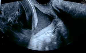
Vasa Praevia: When to Look Closer
Vasa praevia is a near-perfect example of a condition where ultrasound can genuinely change a pregnancy’s outcome. Vasa praevia can be silent; the baby gives no warning, and the only thing standing between a safe, planned birth and a preventable tragedy is early diagnosis in experienced hands.
Patients diagnosed with vasa praevia have emphasised the importance of receiving clear, evidence-based information about the risks and benefits of different care pathways. The primary benefit of screening for vasa praevia is that antenatal diagnosis significantly increases the likelihood of perinatal survival.
This post summarises current best practice, drawing on the RANZCOG guideline Vasa Praevia (C-Obs 47), Version 3 (July 2025), the most relevant evidence-based resource for practice in Australia and Aotearoa New Zealand. Written for referring GPs and clinicians, it covers what vasa praevia is, why it matters so much at delivery, the histories and findings that should raise your index of suspicion, and when targeted specialist assessment is worth arranging. (You may also see it written as vasa previa, the spelling used in much of the international literature; the two terms describe the same condition.)
Table of contents
- What is vasa praevia?
- Why diagnosis matters
- Who is at risk?
- How is vasa praevia diagnosed?
- When to refer to a tertiary unit
- Delivery and management
- Refer your patient to QUFW
What is vasa praevia?
Vasa praevia is the presence of unprotected fetal blood vessels within the amniotic membranes over or near the cervical os. Rupture of the membranes before or during labour may result in fetal death from rapid blood loss if an unprotected fetal blood vessel near the cervix is damaged.
Three types are described:
- Type 1 – characterised by a velamentous insertion of the umbilical cord onto the membranes distant to the placenta, with the vessels traversing the membranes over the cervix before reaching the placenta.
- Type 2 – characterised by a succenturiate, multilobed or bilobed placenta with fetal vessels connecting the lobes overlaying the cervix.
- Type 3 – characterised by abnormal fetal blood vessels branching off the placenta and lying unprotected near the cervical os, arising after resolution of placenta praevia or low-lying placenta.
A systematic review of 24 observational studies reported 1 case of vasa praevia for every 1,271 pregnancies (95% CI 990 to 1,692) (Zhang et al., 2023). Australian data suggest a lower rate as cited in Sullivan et al. (2017) with 63 cases from approximately 294,045 pregnancies, or 1 in 4,673. The lower Australian incidence may be explained in part by low multiple pregnancy rates arising from single embryo transfer policies (RANZCOG, 2025).
Why diagnosis matters
Screening programs for vasa praevia have been shown to be highly sensitive. When vasa praevia is not diagnosed before labour, the estimated perinatal mortality is ==44% to 70% (RANZCOG, 2025)==, and that mortality persists even with an urgent caesarean once a vessel has ruptured. The classic intrapartum presentation is painless dark-red vaginal bleeding after rupture of membranes, accompanied by acute fetal compromise. By the time this happens, the window to act is narrow.
When the diagnosis is made antenatally and a planned caesarean is performed before labour, perinatal survival is 98.6% (95% CI 96.7 to 99.7%), compared with 72.1% (95% CI 50.6 to 89.4%) where no antenatal diagnosis is made (Zhang et al., 2021). Intact perinatal survival, meaning survival without severe impairment, shows the same pattern: 96.7% with an antenatal diagnosis against 28.1% without (Zhang et al., 2021).
In short, the condition is almost entirely survivable if found in time, and frequently fatal if not. That dichotomy is what justifies screening and vigilance.
Who is at risk?
Approximately 83% of women with vasa praevia have at least one identifiable risk factor (RANZCOG, 2025). The strongest ultrasound predictors are a low-lying placenta and a velamentous cord insertion.
Pooled prevalence of each risk factor among vasa praevia cases (Pavalagantharajah et al., 2020):
| Risk factor | Prevalence in vasa praevia cases (95% CI) |
|---|---|
| Low-lying placenta or resolved placenta praevia | 61.5% (53.0–70.0%) |
| Velamentous cord insertion | 52.2% (39.6–64.7%) |
| Bilobed or succenturiate-lobed placenta | 33.3% (20.9–45.7%) |
| Pregnancy through assisted reproductive technology | 28.2% (20.1–36.3%) |
| Multiple gestation | 8.9% (5.3–12.5%) |
Risk factors are common, and most women with them will not have vasa praevia. Velamentous cord insertion occurs in roughly 2% of pregnancies, while vasa praevia affects approximately 0.08%. Risk factors raise the index of suspicion and determine who needs a follow-up scan but they are not the diagnosis itself.
It is also worth noting that findings identified in the second trimester frequently resolve. Vasa praevia resolves in 14% to 39% of cases, with resolution far more likely when the finding is made before 24 to 26 weeks (RANZCOG, 2025). This is precisely why the diagnosis is confirmed later in pregnancy rather than applied at the morphology scan.
How is vasa praevia diagnosed?
Diagnosis is made with ultrasound, and RANZCOG describes a two-stage pathway.
Screening — 20 to 22 week morphology scan. RANZCOG suggests all women be offered screening for vasa praevia at the time of the second trimester anatomy scan (Evidence-based recommendation 1). A good quality report will comment on the location of the placental cord insertion, the morphology of the placenta, and clear documentation of a colour Doppler sweep of the cervix to assess for low-lying fetal vessels. At this stage, no distinction is drawn between vasa praevia and low-lying fetal vessels: any fetal vessels within 50 mm of the internal cervical os are characterised as low-lying fetal vessels.
Diagnosis — transvaginal ultrasound at 28 to 32 weeks. Where any of the following are present at the morphology scan, or where a woman with risk factors has not had a previous scan, the diagnosis should be confirmed by transvaginal ultrasound at 28 to 32 weeks, or earlier if clinically indicated (Evidence-based recommendation 2):
- low-lying fetal vessels (within 50 mm of the cervical os)
- velamentous cord insertion
- low-lying placenta
- bilobed or multilobed placenta
At that third trimester scan, fetal vessels within 20 mm of the cervical os identified by transvaginal ultrasound are diagnostic of vasa praevia (Evidence-based recommendation 3). Vessels between 20 mm and 50 mm are defined as low-lying fetal vessels, and vessels beyond 50 mm require no further investigation for vasa praevia.
Transvaginal ultrasound with Doppler is the preferred approach for evaluating the cervical os and lower uterine segment, providing high resolution of small vessels and a more accurate assessment of the vessel-to-os distance. Where vessels remain within 50 mm at the early third trimester scan, a repeat scan at or after 32 weeks should be considered to confirm their final location.
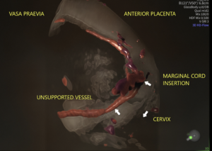
3D Rendered Volume with Colour Doppler
There is no international consensus on the distance that defines vasa praevia. In a 2024 survey of 57 experts, 34% used a 20 mm threshold, 32% used 50 mm, and 21% did not rely on distance at all (RANZCOG, 2025). The RANZCOG thresholds represent a pragmatic position rather than a settled one.
When to refer to a tertiary unit
For GPs and clinicians in shared-care or community settings, the referral triggers are straightforward:
- A suspected or confirmed finding on ultrasound should prompt referral to specialist obstetric care for a confirmatory transvaginal scan at 28 to 32 weeks, and ongoing management planning.
- Suspected vasa praevia, together with clinical features of preterm labour or antepartum haemorrhage occurring before the third-trimester confirmatory scan, warrants consultation with a Maternal Fetal Medicine (MFM) specialist for management of the pregnancy.
- At the extremes of prematurity, or where the diagnosis is uncertain, MFM consultation is again advisable to balance the competing risks.
An equivocal or incomplete view of the cord insertion or the vessels near the os is reasonable grounds to refer for a closer look. The aim is simply to ensure that, where the risk exists, the vessels have been specifically assessed. When vasa praevia is excluded, that is genuine reassurance for the patient and for you. When it is identified, a clear plan can be implemented.
Delivery and management
Once vasa praevia is confirmed, patient management centres on planning a caesarean birth before the onset of labour, while weighing the risks of prematurity against the risk of vessel rupture. Care should be individualised and discussed in the context of patient preferences and individual circumstances.
For asymptomatic women with confirmed vasa praevia, inpatient versus outpatient management from 32+0 weeks should be discussed rather than mandated. Outpatient management is reasonable for asymptomatic women, particularly those without additional risk factors and with good access to emergency services. When weighing this up, consider additional risk factors for preterm labour multiple gestation, a short cervix, or a history of preterm birth. Women with low-lying fetal vessels (20 to 50 mm) can generally be managed as outpatients with ongoing antenatal care and birth planning.
Current guidance on timing of birth, balancing prematurity against the risk of vessel rupture in labour (RANZCOG, 2025):
- Asymptomatic, no preterm-birth risk factors: consider caesarean from 36+0 weeks. Evidence suggests 36+0 to 36+6 weeks may best balance the risks of prematurity against those of vasa praevia complications.
- Risk factors for preterm birth present: consider caesarean from 34+0 weeks.
- Symptomatic (bleeding, preterm labour): consider urgent caesarean, balancing the risks of prematurity against the likelihood of fetal compromise.
Birth planning should be guided by clinical features, patient preferences, and access to specialist obstetric and neonatal services, with the aim of ensuring birth before the onset of labour or complications.
Refer your patient to QUFW
At QUFW, our consultants are subspecialist obstetricians and gynaecologists with extensive experience in high-risk pregnancies. Vasa praevia is uncommon, but primary care providers should offer a routine ultrasound at 20 to 22 weeks (morphology scan) to assess for this condition in all pregnancies.
If you would like to contact us to discuss a patient you would like assessed, or if your patient would like to book an appointment, click here.
References
Pavalagantharajah, S., Villani, L. A., & D’Souza, R. (2020). Vasa previa and associated risk factors: A systematic review and meta-analysis. American Journal of Obstetrics & Gynecology MFM, 2(3), 100117.
Royal Australian and New Zealand College of Obstetricians and Gynaecologists. (2025). Vasa praevia (C-Obs 47, Clinical Guideline Version 3).
Sullivan, E. A., Javid, N., Duncombe, G., Li, Z., Safi, N., Cincotta, R., Homer, C. S. E., Halliday, L., & Oyelese, Y. (2017). Vasa previa diagnosis, clinical practice, and outcomes in Australia. Obstetrics & Gynecology, 130(3), 591–598.
Zhang, W., Geris, S., Al-Emara, N., Ramadan, G., Sotiriadis, A., & Akolekar, R. (2021). Perinatal outcome of pregnancies with prenatal diagnosis of vasa previa: Systematic review and meta-analysis. Ultrasound in Obstetrics & Gynecology, 57(5), 710–719.
Zhang, W., Giacchino, T., Chanyarungrojn, P. A., Ionescu, O., & Akolekar, R. (2023). Incidence of vasa praevia: A systematic review and meta-analysis. BMJ Open, 13(9), e075245.
Content Disclaimer
The information provided on this website is for educational and informational purposes only. It is not intended as a substitute for professional medical advice, diagnosis, or treatment. Always seek the advice of your obstetrician or other qualified provider for any questions you may have regarding a medical condition or treatment, and before undertaking a new healthcare regimen.
The content on this website is not intended to be a comprehensive source of information on any particular topic and should not be relied upon as such. The authors and publishers of this website are not liable for any damages or injury resulting from the use or misuse of the information provided on this website.